Meta-Analysis
. 2015 Dec 15;43(6):1199-211.
doi: 10.1016/j.immuni.2015.11.003.

Integrated, Multi-cohort Analysis Identifies Conserved Transcriptional Signatures across Multiple Respiratory Viruses

Affiliations
Meta-Analysis

Integrated, Multi-cohort Analysis Identifies Conserved Transcriptional Signatures across Multiple Respiratory Viruses

Marta Andres-Terre et al. Immunity. .

Abstract

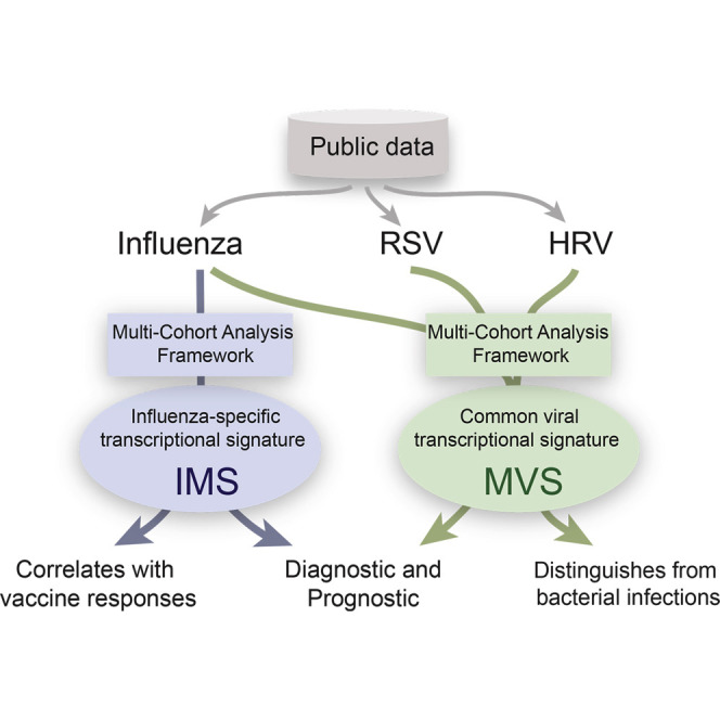
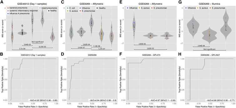
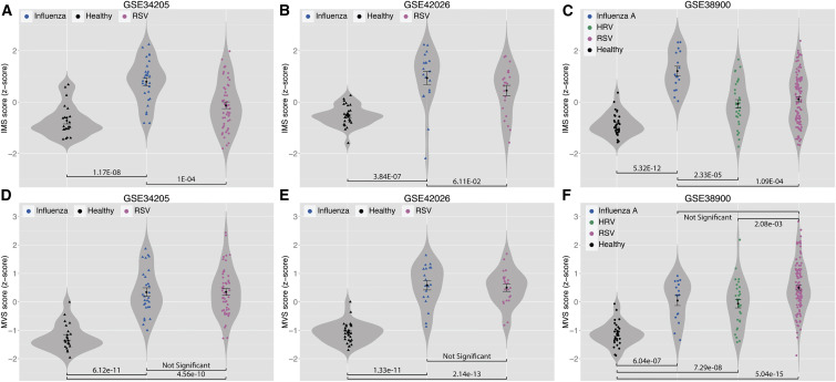
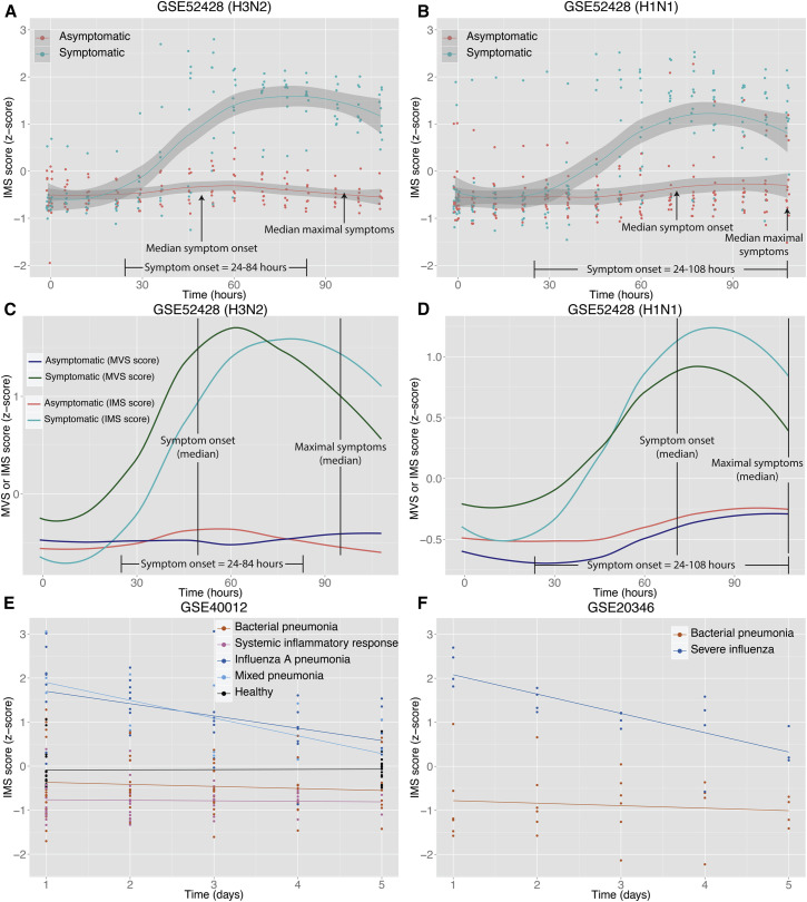
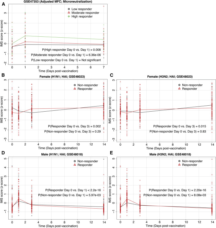
Respiratory viral infections are a significant burden to healthcare worldwide. Many whole genome expression profiles have identified different respiratory viral infection signatures, but these have not translated to clinical practice. Here, we performed two integrated, multi-cohort analyses of publicly available transcriptional data of viral infections. First, we identified a common host signature across different respiratory viral infections that could distinguish (1) individuals with viral infections from healthy controls and from those with bacterial infections, and (2) symptomatic from asymptomatic subjects prior to symptom onset in challenge studies. Second, we identified an influenza-specific host response signature that (1) could distinguish influenza-infected samples from those with bacterial and other respiratory viral infections, (2) was a diagnostic and prognostic marker in influenza-pneumonia patients and influenza challenge studies, and (3) was predictive of response to influenza vaccine. Our results have applications in the diagnosis, prognosis, and identification of drug targets in viral infections.


Figures

None
Graphical abstract
Figure 1
Figure 1
Discovery and Validation of Meta Virus Signature Effect size heatmaps of 396-gene MVS in 5 discovery (A) and 10 validation (B) cohorts. Each column is a gene and row is a cohort. The first row in both heatmaps displays summary effect size for each gene in discovery or validation cohorts. Genes are sorted in decreasing order of their summary effect size in discovery cohorts for both heatmaps.
Figure 2
Figure 2
MVS Scores in Four Independent Cohorts of Blood Samples (A) Comparison of MVS scores in virus-negative, afebrile controls, and patients infected with bacteria or HRV. (B) Comparison of MVS scores in healthy controls and patients infected with SARS coronavirus. Error bars indicate mean ± SE for a given group of samples. Width of a violin plot indicates density of samples, where each dot represents a sample. (C and D) MVS scores in symptomatic and asymptomatic subjects inoculated with influenza (H3N2 in C or H1N1 in D). Smoothed lines indicate loess curves for symptomatic and asymptomatic subjects. Gray bars indicate 95% confidence intervals for each group. See also Figure S1.
Figure 3
Figure 3
11-Gene Influenza Meta Signature (A–K) 11 genes were significantly overexpressed during influenza infection in all discovery cohorts analyzed. The x axes represent standardized mean difference between influenza and control samples, computed as Hedges’ g, in log2 scale. The size of the blue rectangles is inversely proportional to the SEM in the study. Whiskers represent the 95% confidence interval. The orange diamonds represent overall, combined mean difference for a given gene. Width of the diamonds represents the 95% confidence interval of overall mean difference. (L) Network analysis by Ingenuity Pathway Analysis showed that 10 out of the 11 genes are part of a single regulatory network. Blue nodes in the network represent IMS genes, and red nodes represent genes that were significantly overexpressed during influenza infection but are not included in the IMS.
Figure 4
Figure 4
The IMS Discriminates Influenza Infection from Bacterial Infections with High Specificity and Sensitivity in Multiple Cohorts ROCs are for distinguishing patients with influenza infection from all other samples in a given cohort. (A and B) For GSE40012, only samples on the first day of admission are used for violin plot (A) and ROC (B). (C–H) Violin plots (C, E, G) and ROCs (D, F, H) for samples profiled with three microarray platforms in GSE6269 are displayed individually. Error bars indicate mean ± SE for a given group of samples. Width of a violin plot indicates density of samples, where each dot represents a sample. See also Figure S3.
Figure 5
Figure 5
The IMS Scores Distinguish Influenza-Infected Patients from Healthy Controls and Patients with Other Respiratory Viral Infections but the MVS Scores Do Not (A–C) In GSE34205 (A) and GSE42026 (B), samples from influenza patients and healthy controls were used for discovery of the IMS, but samples from RSV-infected patients were not used. None of the samples from GSE38900 (C) were used for discovery of the IMS. (D–F) All samples from GSE34205 (D) and GSE42026 (E) were used for identification of the MVS. None of the samples from GSE38900 (F) were used for identification of MVS. Error bars indicate mean ± SE for a given group of samples. Width of a violin plot indicates density of samples, where each dot represents a sample. See also Figure S3.
Figure 6
Figure 6
IMS Score Is a Prognostic Marker of Influenza Infection (A and B) IMS scores in symptomatic and asymptomatic subjects inoculated with influenza A (H3N2 in A; H1N1 in B). Smoothed lines indicate loess curves for symptomatic and asymptomatic subjects. Gray bars indicate 95% confidence interval. (C and D) Comparison of MVS and IMS scores in the same subjects inoculated with H3N2 (C) or H1N1 (D). (E and F) Change in IMS scores in healthy controls and patients with non-infectious systemic inflammatory response, influenza pneumonia, bacterial pneumonia, or both (influenza and bacterial pneumonia) during their stay in the hospital. See also Figure S4.
Figure 7
Figure 7
IMS Score Increases Significantly in Vaccine Responders (A) Change in IMS scores on day 1 after vaccination for high (p = 0.008), moderate (p = 6.36 × 10−6), and low (p > 0.05) responders defined based on microneutralization titers. (B and C) Change in IMS scores for vaccine responders and non-responders, defined based on HAI titers, in a female cohort after influenza vaccination (H1N1 in B; H3N2 in C). (D and E) Change in IMS scores for vaccine responders and non-responders, defined based on HAI titers, in a male cohort after influenza vaccination (H1N1 in D; H3N2 in E). See also Figure S5.

References

    1. Babcock H.M., Merz L.R., Dubberke E.R., Fraser V.J. Case-control study of clinical features of influenza in hospitalized patients. Infect. Control Hosp. Epidemiol. 2008;29:921–926. - PubMed
    1. Barrett T., Suzek T.O., Troup D.B., Wilhite S.E., Ngau W.-C., Ledoux P., Rudnev D., Lash A.E., Fujibuchi W., Edgar R. NCBI GEO: mining millions of expression profiles--database and tools. Nucleic Acids Res. 2005;33:D562–D566. - PMC - PubMed
    1. Benjamini Y., Hochberg Y. Controlling the false discovery rate: A practical and powerful approach to multiple testing. J. R. Statist. Soc., B. 1995;57:289–300.
    1. Bermejo-Martin J.F., Martin-Loeches I., Rello J., Antón A., Almansa R., Xu L., Lopez-Campos G., Pumarola T., Ran L., Ramirez P. Host adaptive immunity deficiency in severe pandemic influenza. Crit. Care. 2010;14:R167. - PMC - PubMed
    1. Chen R., Khatri P., Mazur P.K., Polin M., Zheng Y., Vaka D., Hoang C.D., Shrager J., Xu Y., Vicent S. A meta-analysis of lung cancer gene expression identifies PTK7 as a survival gene in lung adenocarcinoma. Cancer Res. 2014;74:2892–2902. - PMC - PubMed

Publication types