. 2016 Jan 26;7(4):4490-506.
doi: 10.18632/oncotarget.6606.

Galiellalactone induces cell cycle arrest and apoptosis through the ATM/ATR pathway in prostate cancer cells

Affiliations

Galiellalactone induces cell cycle arrest and apoptosis through the ATM/ATR pathway in prostate cancer cells

Víctor García et al. Oncotarget. .

Abstract

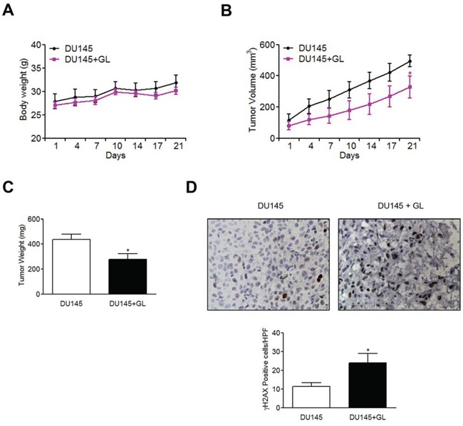
Galiellalactone (GL) is a fungal metabolite that presents antitumor activities on prostate cancer in vitro and in vivo. In this study we show that GL induced cell cycle arrest in G2/M phase, caspase-dependent apoptosis and also affected the microtubule organization and migration ability in DU145 cells. GL did not induce double strand DNA break but activated the ATR and ATM-mediated DNA damage response (DDR) inducing CHK1, H2AX phosphorylation (fH2AX) and CDC25C downregulation. Inhibition of the ATM/ATR activation with caffeine reverted GL-induced G2/M cell cycle arrest, apoptosis and DNA damage measured by fH2AX. In contrast, UCN-01, a CHK1 inhibitor, prevented GL-induced cell cycle arrest but enhanced apoptosis in DU145 cells. Furthermore, we found that GL did not increase the levels of intracellular ROS, but the antioxidant N-acetylcysteine (NAC) completely prevented the effects of GL on fH2AX, G2/M cell cycle arrest and apoptosis. In contrast to NAC, other antioxidants such as ambroxol and EGCG did not interfere with the activity of GL on cell cycle. GL significantly suppressed DU145 xenograft growth in vivo and induced the expression of fH2AX in the tumors. These findings identify for the first time that GL activates DDR in prostate cancer.

Keywords: ATM/ATR; CHK1; cancer; cell cycle; galiellalactone.


Conflict of interest statement

CONFLICTS OF INTEREST

The authors declare no conflict of interest.

Figures

Figure 1
Figure 1. GL induces G2/M phase cell-cycle arrest
A. DU145 cells were exposed to various doses of GL (1, 10 and 20 μM) during 6, 12 or 24 h and cell cycle was analyzed by PI staining and flow cytometry. Representative histograms are shown. B. Quantitation of percentages of the cells in each phase of the cell cycle. Data are the means of three independent experiments ± SD. *P<0.05; **P<0.01; ***P<0.001 compared with the control group. C. Effect of GL (24 h) on cell cycle in human normal dermal fibroblasts and RWPE-1 cells. Representative histograms are shown.
Figure 2
Figure 2. GL induction of cell-cycle arrest is mediated by a caspase-independent pathway
A. DU145 cells were treated with GL in the absence or the presence or the pan-caspase inhibitor Z-VAD-FMK (40 μM) for 48 h and protein expression of PARP and cleavaged caspase-3 was analyzed by immunoblot. B. DU145 cells were treated as above for 48 h, stained with Annexin V and PI and analyzed by flow cytometry. Representative plots and percentages are shown. C. DU145 cells were treated as in A and cell cycle distribution was determined by flow cytometry. Quantitation of percentages of the cells in each phase of the cell cycle. Data are the means of three independent experiments ± SD. ***P<0.001 compared with the control group.
Figure 3
Figure 3. Effect of GL on cell morphology and cytoskeletal structure
A. Double immunofluorescent staining of actin (red) and α-tubulin (green) in DU145 cells treated with cytochalasin D (10 μM), GL (10 μM), nocodazole (100 ng/ml) and docetaxel (10 nM) for 6 h. The nuclei were counterstained with DAPI (blue). Cells were visualized by confocal microscopy (x63). B. Representative cell cycle profiles obtained by FACS at 24 h after the treatment with the indicated compounds.
Figure 4
Figure 4. GL inhibits cell motility
A. DU145 cells were pre-incubated with mitomycin C (5 μg/ml) for 1 h and treated with GL at 10 and 20 μM for 24 h and cell cycle analyzed by PI staining and flow cytometry. Representative histograms are shown. B. DU145 cells were pre-incubated with mitomycin C (5 μg/ml) for 1 h, treated or not with GL at 10 μM for 24 h and relative wound density analyzed at different time points over a period of 24 h. The measurements are from wounds made on a monolayer of DU145 cells cultured in the presence of GL and control. Data are the means of three experiments ± SE. *P<0.05; **P<0.01 compared with the control group. C. Images of wound healing assay were obtained at 0, 12 or 24 h and the blue areas show the initial wound boundaries at 0 h.
Figure 5
Figure 5. Effect of GL on the expression of cell cycle proteins and DNA damage
A. Kinetic analysis on the steady state of proteins involved in G2/M phase. DU145 cells were treated with GL (10 μM) for the indicated times and the expression of the different proteins analyzed by western blots. B. Protein expression of pCHK1, pCHK2 and CDC25C and C. pATR, pATM, and γH2AX was evaluated by immunoblot in cells stimulated with GL for 24 h. D. Alkaline comet assay was performed to determine DNA fragments in DU145 cells treated with either GL (10 μM) or etoposide for 24 h. Representative images of alkaline comet assay and a graph with the tail moment are shown. ***P<0.001 compared with the control group.
Figure 6
Figure 6. GL activates the ATM/ATR/CHK1 pathway
DU145 cells were pre-incubated for 1 h with either UCN-01 (1 μM) or caffeine (10 mM) and then treated with GL 10 μM for 24 h A, D. Representative cell cycle profiles obtained by flow cytometry at 24 h after the treatment with the indicated compounds. B, E. Identification of DNA damage (pCHK1 and γH2AX) and apoptotic (PARP) proteins. C, F. DU145 cells were treated as above for 48 h, stained with Annexin V and PI and analyzed by FACS. Percentages of Annexin V positive cells are shown. Data are the means of three experiments ± SD. *P<0.05; ***P<0.001 compared with the control group. #P< 0.05 compared with GL 10 μM group.
Figure 7
Figure 7. NAC inhibits GL-induced cell cycle arrest and apoptosis in DU145 cells
A. DU145 cells were treated with either GL or TBHP and the generation of intracellular ROS was determined with fluorescence probe DCFH2-DA. ***P<0.001 compared with the positive control group. B. DU145 cells were pre-incubated either NAC (1 mM), epigallocatechin (100 μM) or ambroxol (100 μM) followed by GL 10 μM treatment. Representative cell cycle profiles obtained by FACS after 24 h of treatment are shown. C. Protein expression of PARP, Caspase-3 and γH2AX was determined by western blot. D. DU145 cells were treated as above for 48 h, stained with Annexin V and PI and analyzed by FACS. Percentages of Annexin V positive cells are shown. Data are the means of triplicate experiments ± SD. **P<0.01 compared with the control group. ##P< 0.01 compared with GL 10 μM group.
Figure 8
Figure 8. GL induces H2AX phosphorylation in prostate tumors
Athymic nude-Foxn1nu mice were injected subcutaneously with DU145 cells. After 4 weeks tumors were established and mice were treated every day during 21 days with GL (3 mg/kg) or DMSO (0.1%). A. Body weight in DU145 and GL-treated groups. B. Tumor volume (mm3) was evaluated every 3-4 days using a caliper. C. Tumor weight. D. γH2AX expression in tumor sections of controls and GL-treated mice DU145. Representative images are shown (x40). Quantitation of γH2AX positive cells/HPF (high power field) as shown in the graph. *P<0.05 compared with DU145 group.
Figure 9
Figure 9. Schematic model for the ability of GL to induce cell cycle arrest and apoptosis
GL induces DNA damage and activation of pATM/ATR, which phosphorylates H2AX and CHK1 to induce cell cycle arrest at the G2/M phase by CDC25C inhibition. Activation of pATM/ATR also induces apoptosis through a CHK1-independent pathway.

References

    1. Torre LA, Bray F, Siegel RL, Ferlay J, Lortet-Tieulent J, Jemal A. Global cancer statistics, 2012. CA. 2015;65:87–108. - PubMed
    1. Feldman BJ, Feldman D. The development of androgen-independent prostate cancer. Nature reviews Cancer. 2001;1:34–45. - PubMed
    1. Pienta KJ, Bradley D. Mechanisms underlying the development of androgen-independent prostate cancer. Clinical cancer research. 2006;12:1665–1671. - PubMed
    1. Katzenwadel A, Wolf P. Androgen deprivation of prostate cancer: Leading to a therapeutic dead end. Cancer letters. 2015 - PubMed
    1. Karantanos T, Corn PG, Thompson TC. Prostate cancer progression after androgen deprivation therapy: mechanisms of castrate resistance and novel therapeutic approaches. Oncogene. 2013;32:5501–5511. - PMC - PubMed

Publication types

MeSH terms