. 2016 Feb 12;291(7):3639-47.
doi: 10.1074/jbc.M115.679704. Epub 2015 Dec 18.

Dimethyl Fumarate Inhibits the Nuclear Factor κB Pathway in Breast Cancer Cells by Covalent Modification of p65 Protein

Affiliations

Dimethyl Fumarate Inhibits the Nuclear Factor κB Pathway in Breast Cancer Cells by Covalent Modification of p65 Protein

Irida Kastrati et al. J Biol Chem. .

Abstract

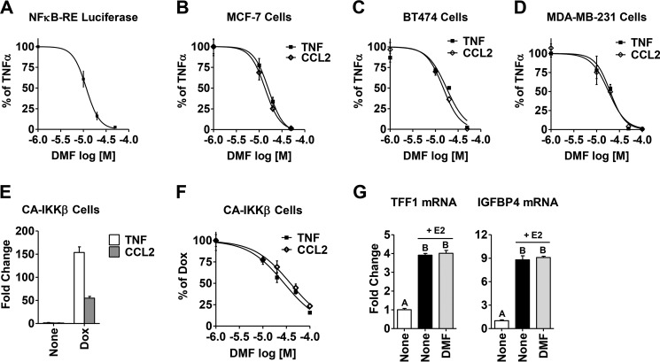
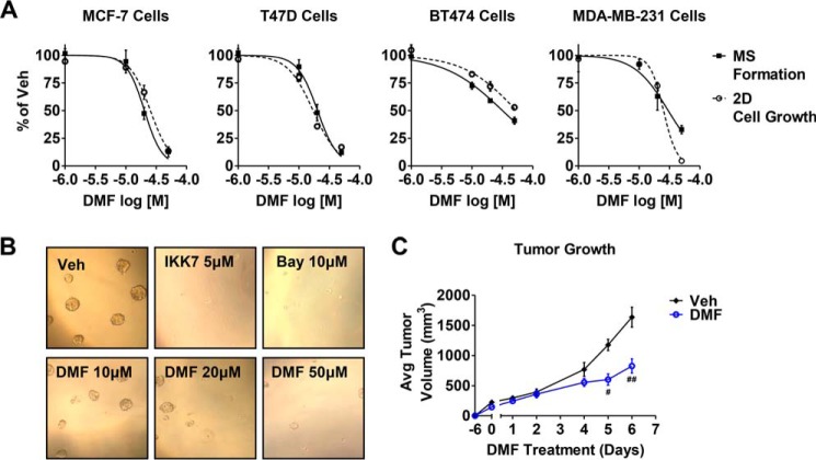
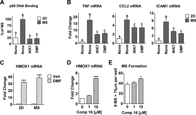
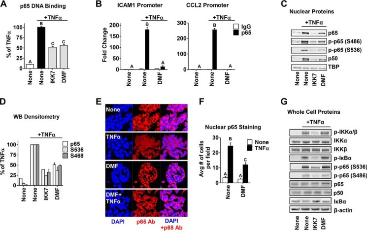
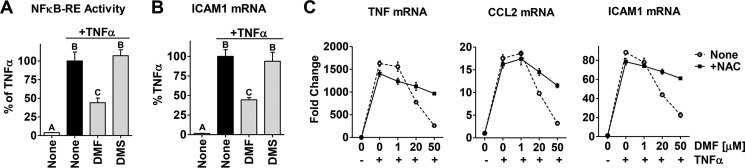
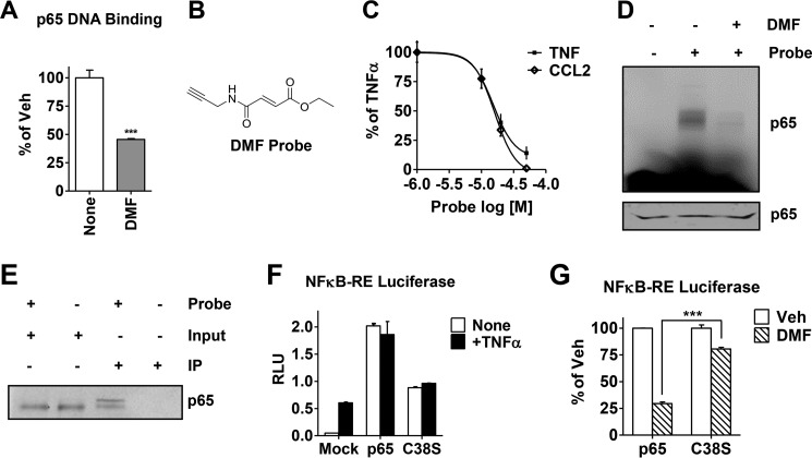
In breast tumors, activation of the nuclear factor κB (NFκB) pathway promotes survival, migration, invasion, angiogenesis, stem cell-like properties, and resistance to therapy--all phenotypes of aggressive disease where therapy options remain limited. Adding an anti-inflammatory/anti-NFκB agent to breast cancer treatment would be beneficial, but no such drug is approved as either a monotherapy or adjuvant therapy. To address this need, we examined whether dimethyl fumarate (DMF), an anti-inflammatory drug already in clinical use for multiple sclerosis, can inhibit the NFκB pathway. We found that DMF effectively blocks NFκB activity in multiple breast cancer cell lines and abrogates NFκB-dependent mammosphere formation, indicating that DMF has anti-cancer stem cell properties. In addition, DMF inhibits cell proliferation and significantly impairs xenograft tumor growth. Mechanistically, DMF prevents p65 nuclear translocation and attenuates its DNA binding activity but has no effect on upstream proteins in the NFκB pathway. Dimethyl succinate, the inactive analog of DMF that lacks the electrophilic double bond of fumarate, is unable to inhibit NFκB activity. Also, the cell-permeable thiol N-acetyl l-cysteine, reverses DMF inhibition of the NFκB pathway, supporting the notion that the electrophile, DMF, acts via covalent modification. To determine whether DMF interacts directly with p65, we synthesized and used a novel chemical probe of DMF by incorporating an alkyne functionality and found that DMF covalently modifies p65, with cysteine 38 being essential for the activity of DMF. These results establish DMF as an NFκB inhibitor with anti-tumor activity that may add therapeutic value in the treatment of aggressive breast cancers.

Keywords: NFκB; breast cancer; chemical modification; covalent modification; cysteine; dimethyl fumarate; drug action; inflammation; inhibitor; mammosphere.


Figures

FIGURE 1.
FIGURE 1.
DMF inhibits TNFα-induced and constitutively active NFκB activity in breast cancer cells. A, NFκB-RE activity was measured in MCF-7 cells following TNFα (10 ng/ml) treatment for 4 h. B–D, expression of the NFκB target genes TNF and CCL2 following TNFα treatment for 2 h was measured by quantitative RT-PCR in MCF-7 cells (B), BT474 cells (C), and MDA-MB-231 cells (D). Increasing concentrations of DMF were added 2 h prior to treatment with TNFα, and the inhibitory activity of DMF is plotted as percent of TNFα alone. E, Dox-induced expression of the NFκB target genes TNF and CCL2, measured by quantitative RT-PCR, is shown in CA-IKKβ cells. F, DMF inhibits expression of the TNF and CCL2 genes in CA-IKKβ cells in a dose-dependent manner. The inhibitory activity of DMF is plotted as percent of Dox alone. IC50 values were calculated with GraphPad software using normalized data. G, DMF has no effect on estrogen receptor target gene TFF1 and IGFBP4 mRNA, measured in MCF-7 cells pretreated with DMF (20 μm) for 2 h, followed by estrogen treatment (E2, 10 nm) for another 2 h. The different letters above bars indicate significant difference between treatments, p < 0.001.
FIGURE 2.
FIGURE 2.
DMF has anti-breast cancer activity both in vitro and in vivo. A, MS formation (solid line) and 2D cell growth (dashed line) in the indicated cell lines were measured after treatment with varying concentrations of DMF. The effect of DMF is plotted as percent of dimethyl sulfoxide vehicle (veh) control. B, representative pictures at ×10 magnification of MCF-7 MS formation upon treatment with DMF or the NFκB inhibitors IKK7 (5 μm) and Bay117082 (Bay, 10 μm). C, effect of DMF (30 mg/kg daily) on MDA-MB-231 xenograft tumor growth is indicated. #, p = 0.0002; ##, p = 0.003. Avg, average.
FIGURE 3.
FIGURE 3.
DMF inhibits the intrinsic NFκB activity in MS culture of breast cancer cells. A, p65 DNA binding activity was measured via ELISA in a conventional adherent 2D culture of MCF-7 cells or MS culture with or without inhibitors (IKK7 1 μm or DMF 50 μm) added for the last 3 h. B, expression of the TNF, CCL2, and ICAM1 genes after 6 h of drug treatment was measured in the same groups described in A. The different letters above the columns indicate significant difference between treatments (p < 0.001). C, DMF (20 μm) up-regulates the expression of HMOX1 mRNA in both 2D and MS of MCF-7 cells. Data are shown as -fold change compared with vehicle (Veh) control. D, compound 16 (Comp 16, 1 and 10 μm) up-regulates the expression of HMOX1 mRNA in MCF-7 cells. E, MS formation is measured in MCF-7 cells treated with compound 16. *, p < 0.01; ***, p < 0.001.
FIGURE 4.
FIGURE 4.
DMF inhibits p65 DNA binding and transcriptional activity and its nuclear translocation in an IKK/IκBα-independent manner. A, p65 DNA binding activity was measured in MCF-7 cells treated with IKK7 (1 μm) or DMF (50 μm) for 2 h, followed by TNFα treatment for 15 min. B, ChIP assays were carried out for p65 and the IgG control following treatment of MCF-7 cells with TNFα for 45 min with or without DMF (50 μm) added 2 h prior to TNFα. The -fold-increase in IgG or p65 occupancy at the ICAM1 (left panel) and CCL2 (right panel) promoters were calculated from the percent input of each pulldown and comparison of each treatment to vehicle controls. C, nuclear extracts of cells treated as in A were prepared, and NFκB transcription factors were examined by Western blotting. Representative Western blotting analyses from three independent experiments are shown. TBP served as a loading control. D, densitometry of nuclear proteins relative to TBP. Data are plotted as percent of TNFα alone. WB, Western blotting. E, representative pictures of IHC staining for nuclear p65 in MCF-7 cells treated with DMF (50 μm) followed by TNFα for 15 min. IHC was performed using an anti-p65 antibody (Ab, red) and DAPI (blue) for nuclear staining and visualized using a Zeiss laser-scanning microscope. F, IHC quantitation of the nuclear p65 content in E. Avg, average. G, whole-cell extracts of cells treated as in A were prepared, and NFκB signaling proteins were examined by Western blotting. Representative Western blotting analyses from three independent experiments are shown. β-Actin served as a loading control. The different letters above the columns in A, B, and E indicate a significant difference between treatments (p < 0.001).
FIGURE 5.
FIGURE 5.
The double bond reactivity of DMF is required to inhibit the NFκB pathway in breast cancer cells. A and B, NFκB-RE activity (A) and ICAM1 (B) gene expression were measure in MCF-7 cells upon treatment with DMF or DMS, 20 μm each, as described in Fig. 1. Data are plotted as percent of TNFα alone. The different letters above the columns indicate a significant difference between treatments (p < 0.001). C, NAC (dashed line) reverses the inhibitory effect of DMF on the TNFα-induced expression of the NFκB target genes TNF, CCL2, and ICAM1 in MCF-7 cells. NAC (500 μm) was added 30 min prior to DMF and TNFα treatment.
FIGURE 6.
FIGURE 6.
DMF covalently modifies p65 both in vitro and in cell lysates, and cysteine 38 is the key residue responsible for DMF activity on the NFκB pathway. A, recombinant p65 DNA binding activity was measured after incubation with DMF (50 μm) for 30 min. Data are plotted as percent vehicle (Veh) control. ***, p < 0.001. B, the chemical structure of the novel alkyne-based DMF probe. C, expression of the NFκB target genes TNF and CCL2 in MCF-7 cells was measured by quantitative RT-PCR upon treatment with varying concentrations of the DMF probe, followed by TNFα. Data are plotted as percent of TNFα alone. D, gel image for labeling of recombinant p65 with the DMF probe (50 μm) measured by in-gel fluorescence. Third lane, DMF (50 μm) was added for 30 min prior to incubation with the DMF probe. Bottom panel, Coomassie staining of the gel indicating equal protein loading. E, biotin IP was carried out in MDA-MB-231 cell lysates cross-linked in the presence or absence of the DMF probe (50 μm). Total p65 protein was then immunoblotted and compared between IPs and inputs (10% of the protein lysate load). F, NFκB-RE activity (shown in relative luciferase units, RLU) was measured in MCF-7 cells transfected with wild-type p65 or C38S-p65 or mock-transfected and then treated with or without TNFα for 4 h. G, NFκB-RE activity was measured in MCF-7 cells transfected with wild-type p65 or C38S-p65 and treated with DMF (50 μm). Data are presented as percent inhibition relative to the vehicle (Veh) control. ***, p < 0.001.

References

    1. Hanahan D., and Weinberg R. A. (2011) Hallmarks of cancer: the next generation. Cell 144, 646–674 - PubMed
    1. Baumgarten S. C., and Frasor J. (2012) Inflammation: an instigator of more aggressive estrogen receptor (ER) positive breast cancers. Mol. Endocrinol. 26, 360–371 - PMC - PubMed
    1. Karin M., Cao Y., Greten F. R., and Li Z. W. (2002) NF-κB in cancer: from innocent bystander to major culprit. Nat. Rev. Cancer 2, 301–310 - PubMed
    1. Kim H. J., Hawke N., and Baldwin A. S. (2006) NF-κB and IKK as therapeutic targets in cancer. Cell Death Differ. 13, 738–747 - PubMed
    1. Nakshatri H., Bhat-Nakshatri P., Martin D. A., Goulet R. J. Jr., and Sledge G. W. Jr. (1997) Constitutive activation of NF-κB during progression of breast cancer to hormone-independent growth. Mol. Cell. Biol. 17, 3629–3639 - PMC - PubMed

Publication types

MeSH terms