

Renal Graft Fibrosis and Inflammation Quantification by an Automated Fourier-Transform Infrared Imaging Technique
- PMID: 26683669
- PMCID: PMC4978043
- DOI: 10.1681/ASN.2015050601
Renal Graft Fibrosis and Inflammation Quantification by an Automated Fourier-Transform Infrared Imaging Technique
Abstract
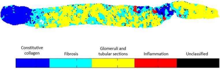
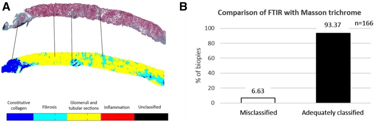
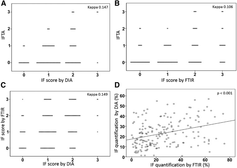
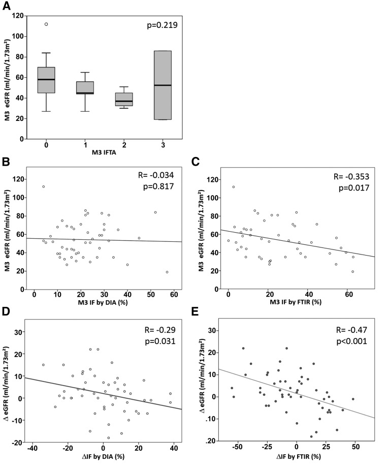
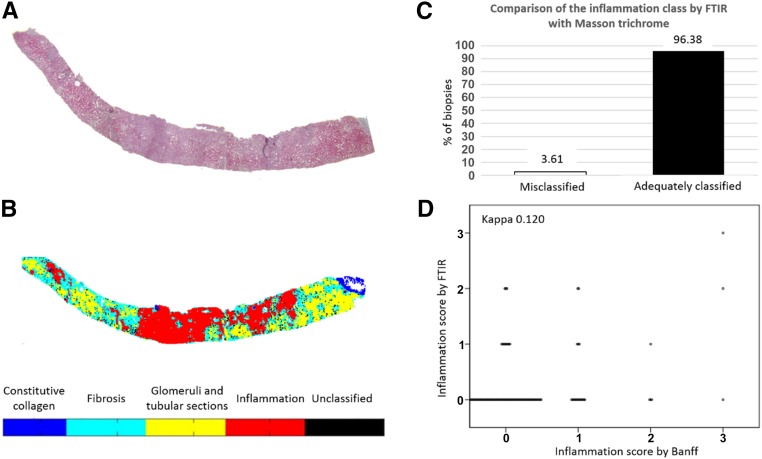
Renal interstitial fibrosis and interstitial active inflammation are the main histologic features of renal allograft biopsy specimens. Fibrosis is currently assessed by semiquantitative subjective analysis, and color image analysis has been developed to improve the reliability and repeatability of this evaluation. However, these techniques fail to distinguish fibrosis from constitutive collagen or active inflammation. We developed an automatic, reproducible Fourier-transform infrared (FTIR) imaging-based technique for simultaneous quantification of fibrosis and inflammation in renal allograft biopsy specimens. We generated and validated a classification model using 49 renal biopsy specimens and subsequently tested the robustness of this classification algorithm on 166 renal grafts. Finally, we explored the clinical relevance of fibrosis quantification using FTIR imaging by comparing results with renal function at 3 months after transplantation (M3) and the variation of renal function between M3 and M12. We showed excellent robustness for fibrosis and inflammation classification, with >90% of renal biopsy specimens adequately classified by FTIR imaging. Finally, fibrosis quantification by FTIR imaging correlated with renal function at M3, and the variation in fibrosis between M3 and M12 correlated well with the variation in renal function over the same period. This study shows that FTIR-based analysis of renal graft biopsy specimens is a reproducible and reliable label-free technique for quantifying fibrosis and active inflammation. This technique seems to be more relevant than digital image analysis and promising for both research studies and routine clinical practice.
Keywords: acute rejection; chronic allograft nephropathy; histopathology; interstitial fibrosis; renal pathology; renal transplantation.
Copyright © 2016 by the American Society of Nephrology.
Figures
Similar articles
-
New computerized color image analysis for the quantification of interstitial fibrosis in renal transplantation.Transplantation. 2011 Oct 27;92(8):890-9. doi: 10.1097/TP.0b013e31822d879a. Transplantation. 2011. PMID: 21926945
-
ARFI-based tissue elasticity quantification and kidney graft dysfunction: first clinical experiences.Clin Hemorheol Microcirc. 2011;49(1-4):527-35. doi: 10.3233/CH-2011-1503. Clin Hemorheol Microcirc. 2011. PMID: 22214724 Clinical Trial.
-
[Recent approaches of quantification of interstitial fibrosis in renal transplantation].Med Sci (Paris). 2009 Nov;25(11):945-50. doi: 10.1051/medsci/20092511945. Med Sci (Paris). 2009. PMID: 19951669 Review. French.
-
Clinical and pathological analyses of interstitial fibrosis and tubular atrophy cases after kidney transplantation.Nephrology (Carlton). 2016 Jul;21 Suppl 1:26-30. doi: 10.1111/nep.12766. Nephrology (Carlton). 2016. PMID: 26972969
-
Chronic allograft nephropathy or interstitial fibrosis and tubular atrophy: what is in a name?Curr Opin Nephrol Hypertens. 2014 May;23(3):245-50. doi: 10.1097/01.mnh.0000444811.26884.2d. Curr Opin Nephrol Hypertens. 2014. PMID: 24626060 Review.
Cited by
-
Artificial Intelligence-Assisted Renal Pathology: Advances and Prospects.J Clin Med. 2022 Aug 22;11(16):4918. doi: 10.3390/jcm11164918. J Clin Med. 2022. PMID: 36013157 Free PMC article. Review.
-
Banff Digital Pathology Working Group: Going digital in transplant pathology.Am J Transplant. 2020 Sep;20(9):2392-2399. doi: 10.1111/ajt.15850. Epub 2020 Apr 19. Am J Transplant. 2020. PMID: 32185875 Free PMC article.
-
Image Analysis Pipeline for Renal Allograft Evaluation and Fibrosis Quantification.Kidney Int Rep. 2021 Apr 24;6(7):1878-1887. doi: 10.1016/j.ekir.2021.04.019. eCollection 2021 Jul. Kidney Int Rep. 2021. PMID: 34307982 Free PMC article.
-
Extracellular Vesicles and Their Relationship with the Heart-Kidney Axis, Uremia and Peritoneal Dialysis.Toxins (Basel). 2021 Nov 4;13(11):778. doi: 10.3390/toxins13110778. Toxins (Basel). 2021. PMID: 34822562 Free PMC article. Review.
-
Label Free Detection of Sensitive Mid-Infrared Biomarkers of Glomerulonephritis in Urine Using Fourier Transform Infrared Spectroscopy.Sci Rep. 2017 Jul 4;7(1):4601. doi: 10.1038/s41598-017-04774-7. Sci Rep. 2017. PMID: 28676642 Free PMC article.
References
-
- Nankivell BJ, Borrows RJ, Fung CL, O’Connell PJ, Allen RD, Chapman JR: The natural history of chronic allograft nephropathy. N Engl J Med 349: 2326–2333, 2003 - PubMed
-
- Nankivell BJ, Borrows RJ, Fung CL, O’Connell PJ, Chapman JR, Allen RD: Calcineurin inhibitor nephrotoxicity: Longitudinal assessment by protocol histology. Transplantation 78: 557–565, 2004 - PubMed
-
- Furness PN, Taub N Convergence of European Renal Transplant Pathology Assessment Procedures (CERTPAP) Project : International variation in the interpretation of renal transplant biopsies: Report of the CERTPAP Project. Kidney Int 60: 1998–2012, 2001 - PubMed
-
- Pape L, Henne T, Offner G, Strehlau J, Ehrich JH, Mengel M, Grimm PC: Computer-assisted quantification of fibrosis in chronic allograft nephropaty by picosirius red-staining: A new tool for predicting long-term graft function. Transplantation 76: 955–958, 2003 - PubMed
-
- Grimm PC, Nickerson P, Gough J, McKenna R, Stern E, Jeffery J, Rush DN: Computerized image analysis of Sirius Red-stained renal allograft biopsies as a surrogate marker to predict long-term allograft function. J Am Soc Nephrol 14: 1662–1668, 2003 - PubMed
MeSH terms
LinkOut - more resources
Full Text Sources
Medical
