

Efficient Small Blob Detection Based on Local Convexity, Intensity and Shape Information
- PMID: 26685229
- PMCID: PMC6991892
- DOI: 10.1109/TMI.2015.2509463
Efficient Small Blob Detection Based on Local Convexity, Intensity and Shape Information
Abstract
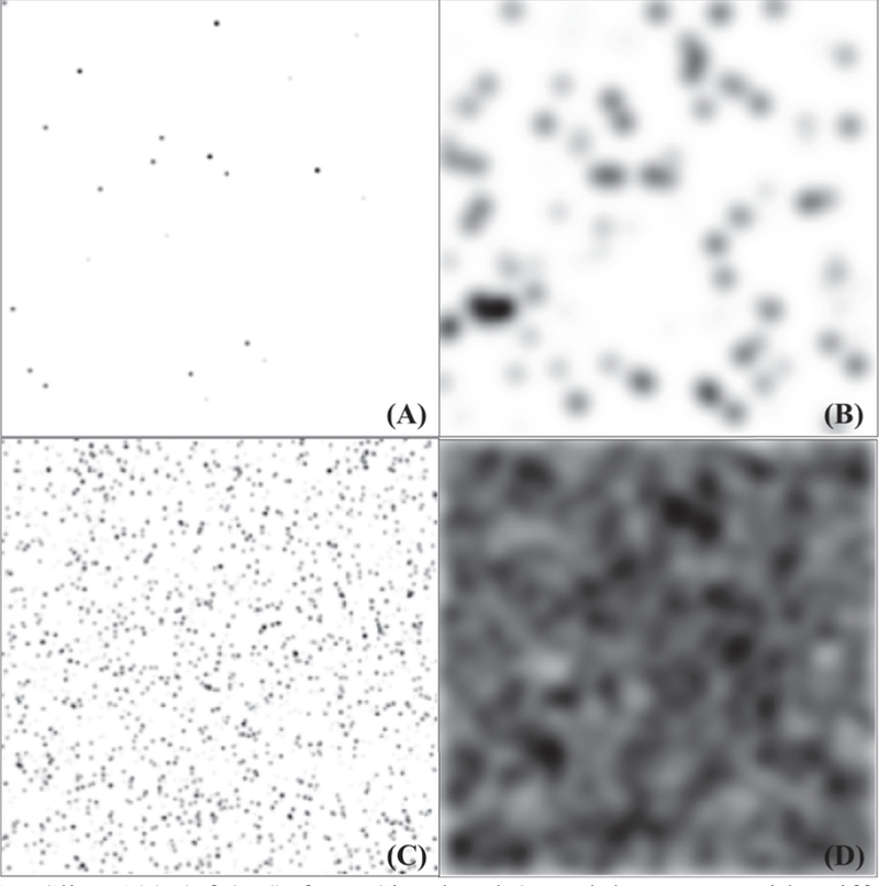
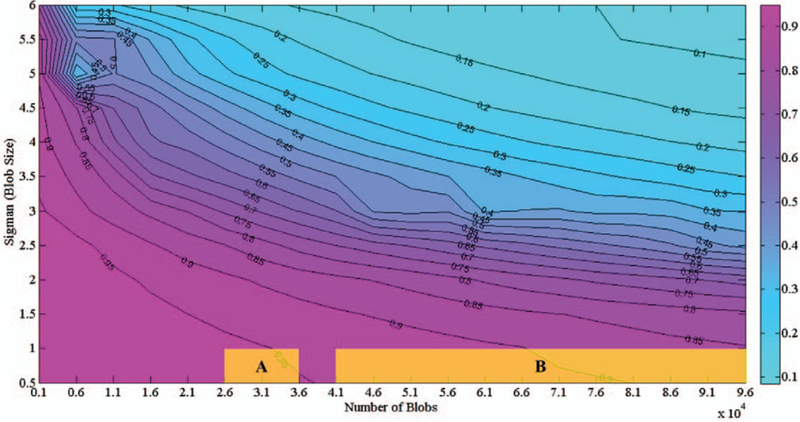
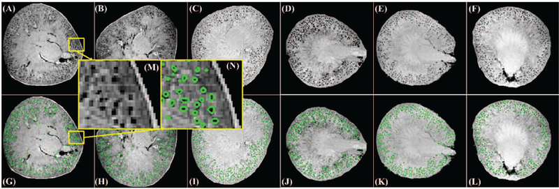
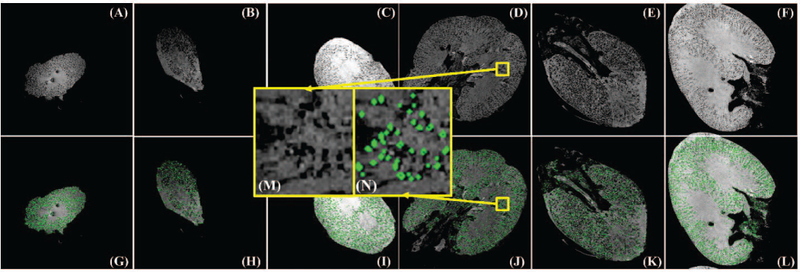
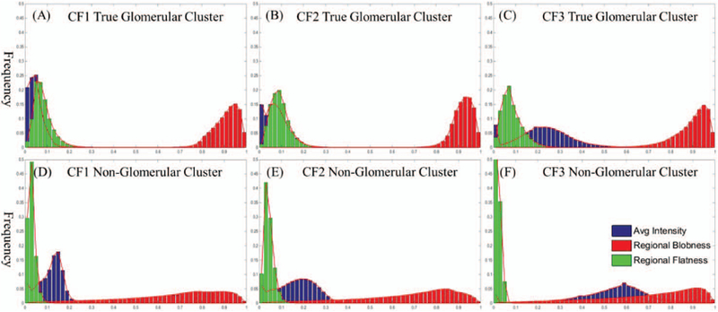
The identification of small structures (blobs) from medical images to quantify clinically relevant features, such as size and shape, is important in many medical applications. One particular application explored here is the automated detection of kidney glomeruli after targeted contrast enhancement and magnetic resonance imaging. We propose a computationally efficient algorithm, termed the Hessian-based Difference of Gaussians (HDoG), to segment small blobs (e.g., glomeruli from kidney) from 3D medical images based on local convexity, intensity and shape information. The image is first smoothed and pre-segmented into small blob candidate regions based on local convexity. Two novel 3D regional features (regional blobness and regional flatness) are then extracted from the candidate regions. Together with regional intensity, the three features are used in an unsupervised learning algorithm for auto post-pruning. HDoG is first validated in a 2D form and compared with other three blob detectors from literature, which are generally for 2D images only. To test the detectability of blobs from 3D images, 240 sets of simulated images are rendered for scenarios mimicking the renal nephron distribution observed in contrast-enhanced, 3D MRI. The results show a satisfactory performance of HDoG in detecting large numbers of small blobs. Two sets of real kidney 3D MR images (6 rats, 3 human) are then used to validate the applicability of HDoG for glomeruli detection. By comparing MRI to stereological measurements, we verify that HDoG is a robust and efficient unsupervised technique for 3D blobs segmentation.
Figures


Similar articles
-
3D small structure detection in medical image using texture analysis.Annu Int Conf IEEE Eng Med Biol Soc. 2016 Aug;2016:6433-6436. doi: 10.1109/EMBC.2016.7592201. Annu Int Conf IEEE Eng Med Biol Soc. 2016. PMID: 28269719
-
Small blob identification in medical images using regional features from optimum scale.IEEE Trans Biomed Eng. 2015 Apr;62(4):1051-62. doi: 10.1109/TBME.2014.2360154. IEEE Trans Biomed Eng. 2015. PMID: 25265624
-
Small Blob Detector Using Bi-Threshold Constrained Adaptive Scales.IEEE Trans Biomed Eng. 2021 Sep;68(9):2654-2665. doi: 10.1109/TBME.2020.3046252. Epub 2021 Aug 23. IEEE Trans Biomed Eng. 2021. PMID: 33347401 Free PMC article.
-
Multivariate statistical model for 3D image segmentation with application to medical images.J Digit Imaging. 2003 Dec;16(4):365-77. doi: 10.1007/s10278-003-1664-9. Epub 2004 Feb 2. J Digit Imaging. 2003. PMID: 14752607 Free PMC article. Review.
-
VoxResNet: Deep voxelwise residual networks for brain segmentation from 3D MR images.Neuroimage. 2018 Apr 15;170:446-455. doi: 10.1016/j.neuroimage.2017.04.041. Epub 2017 Apr 23. Neuroimage. 2018. PMID: 28445774 Review.
Cited by
-
MRI tools for assessment of microstructure and nephron function of the kidney.Am J Physiol Renal Physiol. 2016 Dec 1;311(6):F1109-F1124. doi: 10.1152/ajprenal.00134.2016. Epub 2016 Sep 14. Am J Physiol Renal Physiol. 2016. PMID: 27630064 Free PMC article. Review.
-
In vivo measurements of kidney glomerular number and size in healthy and Os/+ mice using MRI.Am J Physiol Renal Physiol. 2019 Oct 1;317(4):F865-F873. doi: 10.1152/ajprenal.00078.2019. Epub 2019 Jul 24. Am J Physiol Renal Physiol. 2019. PMID: 31339774 Free PMC article.
-
The Hessian Blob Algorithm: Precise Particle Detection in Atomic Force Microscopy Imagery.Sci Rep. 2018 Jan 17;8(1):978. doi: 10.1038/s41598-018-19379-x. Sci Rep. 2018. PMID: 29343783 Free PMC article.
-
Pantograph Detection Algorithm with Complex Background and External Disturbances.Sensors (Basel). 2022 Nov 2;22(21):8425. doi: 10.3390/s22218425. Sensors (Basel). 2022. PMID: 36366124 Free PMC article.
-
Comparative Dissemination of Aerosol and Splatter Using Suction Device during Ultrasonic Scaling: A Pilot Study.Dent J (Basel). 2022 Aug 1;10(8):142. doi: 10.3390/dj10080142. Dent J (Basel). 2022. PMID: 36005240 Free PMC article.
References
-
- Zhang M, Wu T, and Bennett K, “Small Blob Identification in Medical Images Using Regional Features from Optimum Scale,” IEEE Trans Biomed Eng, September 25 2014. - PubMed
-
- Mills PH, Hitchens TK, Foley LM, Link T, Ye Q, Weiss CR, Thompson JD, Gilson WD, Arepally A, Melick JA, Kochanek PM, Ho C, Bulte JW, and Ahrens ET, “Automated detection and characterization of SPIO-labeled cells and capsules using magnetic field perturbations,” Magn Reson Med, vol. 67, pp. 278–89, January 2012. - PMC - PubMed
-
- Sanchez CI, Niemeijer M, Isgum I, Dumitrescu A, Suttorp- Schulten MS, Abramoff MD, and van Ginneken B, “Contextual computer-aided detection: improving bright lesion detection in retinal images and coronary calcification identification in CT scans,” Med Image Anal, vol. 16, pp. 50–62, January 2012. - PubMed
-
- Moon WK, Shen Y-W, Bae MS, Huang C-S, Chen J-H, and Chang R-F, “Computer-aided tumor detection based on multi-scale blob detection algorithm in automated breast ultrasound images,” Medical Imaging, IEEE Transactions on, vol. 32, pp. 1191–1200, 2013. - PubMed
Publication types
MeSH terms
Grants and funding
LinkOut - more resources
Full Text Sources
Other Literature Sources
