. 2016 Jun;124(6):776-84.
doi: 10.1289/ehp.1409627. Epub 2015 Dec 18.

Effects of Chrysotile Exposure in Human Bronchial Epithelial Cells: Insights into the Pathogenic Mechanisms of Asbestos-Related Diseases

Affiliations

Effects of Chrysotile Exposure in Human Bronchial Epithelial Cells: Insights into the Pathogenic Mechanisms of Asbestos-Related Diseases

Giulia Rossana Gulino et al. Environ Health Perspect. 2016 Jun.

Abstract

Background: Chrysotile asbestos accounts for > 90% of the asbestos used worldwide, and exposure is associated with asbestosis (asbestos-related fibrosis) and other malignancies; however, the molecular mechanisms involved are not fully understood. A common pathogenic mechanism for these malignancies is represented by epithelial-mesenchymal transition (EMT), through which epithelial cells undergo a morphological transformation to assume a mesenchymal phenotype. In the present work, we propose that chrysotile asbestos induces EMT through a mechanism involving a signaling pathway mediated by tranforming growth factor beta (TGF-β).

Objectives: We investigated the role of chrysotile asbestos in inducing EMT in order to elucidate the molecular mechanisms involved in this event.

Methods: Human bronchial epithelial cells (BEAS-2B) were incubated with 1 μg/cm2 chrysotile asbestos for ≤ 72 hr, and several markers of EMT were investigated. Experiments with specific inhibitors for TGF-β, glycogen synthase kinase-3β (GSK-3β), and Akt were performed to confirm their involvement in asbestos-induced EMT. Real-time polymerase chain reaction (PCR), Western blotting, and gelatin zymography were performed to detect mRNA and protein level changes for these markers.

Results: Chrysotile asbestos activated a TGF-β-mediated signaling pathway, implicating the contributions of Akt, GSK-3β, and SNAIL-1. The activation of this pathway in BEAS-2B cells was associated with a decrease in epithelial markers (E-cadherin and β-catenin) and an increase in mesenchymal markers (α-smooth muscle actin, vimentin, metalloproteinases, and fibronectin).

Conclusions: Our findings suggest that chrysotile asbestos induces EMT, a common event in asbestos-related diseases, at least in part by eliciting the TGF-β-mediated Akt/GSK-3β/SNAIL-1 pathway.

Citation: Gulino GR, Polimeni M, Prato M, Gazzano E, Kopecka J, Colombatto S, Ghigo D, Aldieri E. 2016. Effects of chrysotile exposure in human bronchial epithelial cells: insights into the pathogenic mechanisms of asbestos-related diseases. Environ Health Perspect 124:776-784; http://dx.doi.org/10.1289/ehp.1409627.


Conflict of interest statement

This paper is dedicated to the memory of D.G., who passed away on 7 October 2015. We all remember our friend as a master in life and science, and we hope he will continue to help us from the place where he is now.

The authors declare they have no actual or potential competing financial interests.

Figures
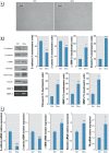
Figure 1
Figure 1
Effects of chrysotile asbestos on cell morphology and epithelial–mesenchymal transition (EMT) marker protein levels in BEAS-2B cells. BEAS-2B cells were cultured for 72 hr without (Ctrl) or with 1 μg/cm2 chrysotile (Chry). (A) Representative microscopy images are shown (10×; scale bar = 40 μm). (B) Expression of epithelial (E-cadherin, β-catenin) and mesenchymal (α-SMA, vimentin, fibronectin) markers checked by Western blotting and evaluation of MMP-2 and MMP-9 activity by zymography. Tubulin was used as a loading control. The image is representative of three independent experiments that produced similar results. Densitometry data are presented as the percent decrease or increase in the protein levelsversus therespective control. Significance versus the respective control: *< 0.005; **< 0.001; ***< 0.0001. (C) Relative gene expression of E-cadherin, β-catenin, α-SMA, vimentin, fibronectin (FN) evaluated by quantitative real-time polymerase chain reaction (qRT-PCR). Data are expressed in units of relative mRNA expression compared with control cells (n = 3). Significance versus the respective control: *< 0.02; **< 0.001; ***< 0.0001.
Figure 2
Figure 2
Effect of crocidolite exposure on cell morphology and alterations in proteins involved in epithelial–mesenchymal transition (EMT) in BEAS-2B cells. BEAS-2B cells were cultured for 7 days without (Ctrl) or with 5 μg/cm2 crocidolite (CROC). (A) Representative microscopy images are shown (10×; scale bar = 40 μm). (B,C) Expression of epithelial (E-cadherin, β-catenin) and mesenchymal (α-SMA, vimentin) markers checked by Western blotting. Tubulin was used as a loading control. The image is representative of three independent experiments that produced similar results. Densitometry data are presented as the percent decrease or percent increase in the protein levels versus the respective control. Significance versus the respective control: *< 0.0001.
Figure 3
Figure 3
TGF-β secretion and neutralizing TGF-β antibody effect in BEAS-2B cells exposed to chrysotile. (A) BEAS-2B cells were incubated in the absence (blue squares) or presence (white squares) of 1 μg/cm2 chrysotile for 30 min and 1, 3, 6 and 72 hr. Afterwards, the supernatants were collected, and TGF-β levels were detected using an ELISA kit. Data are shown as the mean ± SEM (n = 3). TGF-β levels are reported as picograms per milligram of intracellular protein. Significance versus the respective control: *< 0.05; **< 0.02. (B,C) BEAS-2B cells were incubated without (Ctrl) or with 1 μg/cm2 chrysotile (Chry) or with chrysotile and 5 ng/mL of neutralizing anti–TGF-β antibody for 72 hr (Chry + Ab). The expression of epithelial (E-cadherin, β-catenin) and mesenchymal (α-SMA, vimentin) markers was determined by Western blotting. Tubulin was used as a loading control. The image is representative of three independent experiments. Densitometry data are presented as the percent decrease or increase versus control cells. Significance versus the respective control: *< 0.005; **< 0.001; ***< 0.0001. Significance versus chrysotile: °< 0.005; °°< 0.001; °°°< 0.0001. No significant differences were detected for cells coincubated with chrysotile and TGF-β antibody compared with control cells.
Figure 4
Figure 4
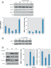
Evaluation of the role of the GSK-3β/SNAIL-1 pathway in E-cadherin gene modulation in BEAS-2B cells. The images are representative of three independent experiments that produced similar results. (A) BEAS-2B cells were incubated in the absence (Ctrl, 6 hr) or presence of 1 μg/cm2 chrysotile for 30 min and 1, 3 and 6 hr (top panel). The expression of phosphorylated GSK-3β (p-GSK-3β) and the accumulation of SNAIL-1 in the nuclei of BEAS-2B cells were examined by Western blotting. p-GSK-3β to GSK-3β ratio values are shown (bottom panel, left). Densitometry data concerning SNAIL-1 accumulation in the nuclei are presented as the percent decrease or increase in the protein levels versus control (Ctrl, 6 hr) (bottom panel, right). Significance versus control (Ctrl, 6 hr): *< 0.0001. (B) BEAS-2B cells were treated without (Ctrl, 6 hr) or with chrysotile (Chry, 1 μg/cm2) together with the specific GSK-3β inhibitor SB216763 (5 μM) for 30 min and 1, 3 and 6 hr. Tubulin and TATA-binding protein (TBP) were used as loading controls for the cytosol and the nucleus, respectively. (C) BEAS-2B cells were treated for 72 hr without (Ctrl) or with chrysotile (1 μg/cm2) in the absence (Chry) or presence (Chry + SB) of SB216763 (5 μM) (left panel). Experiments were performed in triplicate, and densitometry data are presented as the percent decrease or increase in the protein levels vesus the respective control (right panel). Significance versus the respective control: *< 0.005; **< 0.0001. Significance versus chrysotile: °< 0.001.
Figure 5
Figure 5
Evaluation of Akt involvement in GSK-3β regulation in BEAS-2B cells. The images are representative of three independent experiments that produced similar results. (A) BEAS-2B cells were incubated in the absence (Ctrl, 6 hr) or presence of chrysotile (1 μg/cm2) from 30 min up to 6 hr (left panel). Differences in the p-Akt (phosphorylated Akt) to Akt ratio are shown in the right panel. Significance versus control: *< 0.0001. (B) BEAS-2B cells were treated without (Ctrl) or with chrysotile (Chry, 1 μg/cm2) together with the Akt inhibitor (5 μM) from 30 min up to 6 hr. p-GSK-3β = phosphorylated GSK-3β. Tubulin and TATA-binding protein (TBP) were used as loading controls. Differences in the p-GSK-3β to GSK-3β ratio and densitometry data as the percent increase or decrease in SNAIL-1 levels are presented in the right panel. (C) BEAS-2B cells were treated for 72 hr without (Ctrl) or with chrysotile alone (Chry, 1 μg/cm2) or together with 5 μM Akt inhibitor (left panel). Tubulin and TBP were used as loading controls. Densitometry data are presented as the percent decrease or increase in the protein levels versus the respective control (right panel). With regard to the SNAIL-1 densitometry data, no significant differences were detected for cells coincubated with chrysotile and Akt inhibitor compared with control cells. Significance versus the respective control: *< 0.0001. Significance versus chrysotile: °< 0.0001.
Figure 6
Figure 6
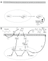
Selected mechanisms potentially involved in TGF-β–mediated epithelial–mesenchymal transition (EMT). (A) Under normal conditions, GSK-3β-mediated phosphorylation of nuclear SNAIL-1 allows its nuclear export and subsequent cytosolic degradation (Zhou et al. 2004). (B) In our study on BEAS-2B cells, chrysotile exposure induced early production of ROS (1), which we hypothesize subsequently activates the latent form of TGF-β (2). Upon release, TGF-β is stabilized and directly presented to its receptors, which then associate and activate a variety of signaling pathways. Both Smad-mediated (3) and non–Smad-mediated (6) pathways are involved (Peinado et al. 2003; Xie et al. 2014). Increased levels of TGF-β result in the activation of the Smad-mediated pathway, suppression of epithelial genes (e.g., E-cadherin), and induction of EMT mesenchymal markers (4) through activation/induction of and coassociation with a variety of transcription factors (including SNAIL-1) (5). We hypothesize that the non–Smad-mediated pathway leads to Akt activation (6) and GSK-3β inactivation (7), and induction of SNAIL-1 nuclear stabilization and genes targeting promotion (8), thus contributing to EMT. In addition, we hypothesize that MMPs secreted by cells (9) may promote the late activation of TGF-β (10), resulting in potentially continuous autocrine cell stimulation that may further reinforce signaling for EMT promotion (11).

References

    1. Acloque H, Adams MS, Fishwick K, Bronner-Fraser M, Nieto MA. Epithelial-mesenchymal transitions: the importance of changing cell state in development and disease. J Clin Invest. 2009;119(6):1438–1449. - PMC - PubMed
    1. Barrallo-Gimeno A, Nieto MA. The Snail genes as inducers of cell movement and survival: implications in development and cancer. Development. 2005;132(14):3151–3161. - PubMed
    1. Bass DA, Parce JW, Dechatelet LR, Szejda P, Seeds MC, Thomas M. Flow cytometric studies of oxidative product formation by neutrophils: a graded response to membrane stimulation. J Immunol. 1983;130(4):1910–1917. - PubMed
    1. Batra H, Antony VB. Pleural mesothelial cells in pleural and lung diseases. J Thorac Dis. 2015;7(6):964–980. - PMC - PubMed
    1. Bhowmick NA, Ghiassi M, Bakin A, Aakre M, Lundquist CA, Engel ME, et al. Transforming growth factor-β1 mediates epithelial to mesenchymal transdifferentiation through a RhoA-dependent mechanism. Mol Biol Cell. 2001;12(1):27–36. - PMC - PubMed

Publication types

MeSH terms

Substances