Review
. 2016 May;1863(5):934-55.
doi: 10.1016/j.bbamcr.2015.12.005. Epub 2015 Dec 11.

Peroxisomes in brain development and function

Affiliations
Review

Peroxisomes in brain development and function

Johannes Berger et al. Biochim Biophys Acta. 2016 May.

Abstract

Peroxisomes contain numerous enzymatic activities that are important for mammalian physiology. Patients lacking either all peroxisomal functions or a single enzyme or transporter function typically develop severe neurological deficits, which originate from aberrant development of the brain, demyelination and loss of axonal integrity, neuroinflammation or other neurodegenerative processes. Whilst correlating peroxisomal properties with a compilation of pathologies observed in human patients and mouse models lacking all or individual peroxisomal functions, we discuss the importance of peroxisomal metabolites and tissue- and cell type-specific contributions to the observed brain pathologies. This enables us to deconstruct the local and systemic contribution of individual metabolic pathways to specific brain functions. We also review the recently discovered variability of pathological symptoms in cases with unexpectedly mild presentation of peroxisome biogenesis disorders. Finally, we explore the emerging evidence linking peroxisomes to more common neurological disorders such as Alzheimer's disease, autism and amyotrophic lateral sclerosis.

Keywords: D-bifunctional protein deficiency; Lipid metabolism; Plasmalogen; Rhizomelic chondrodysplasia punctata; X-linked adrenoleukodystrophy; Zellweger spectrum disorder.


Figures

Fig.1
Fig.1
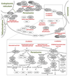
Schematic drawing linking peroxisomal disease-related proteins to individual metabolic pathways. Upper part: Proteins are grouped according to their function in biosynthetic or degradative metabolic pathways, ROS homeostasis, proteolytic activity, transport of metabolites across the peroxisomal membrane (ABCD and PMP proteins), and the import of matrix and membrane proteins (PEX proteins). Ovals represent proteins that are involved in peroxisomal functions (not complete); gray ovals, proteins for which mutations have been linked to a human disease (for full name see Table 1). The degradation of various fatty acids and bile acid precursors is symbolized by the frame depicting the homodimeric transporters (ABCD1–3) and the terms α- and β-oxidation, illustrated in more detail below. Lower part: Proteins are grouped into the degradation pathways for different activated fatty acids (fatty acyl-CoA: saturated, unsaturated, dicarboxylic, branched-chain) and the side chain shortening of di- and trihydroxycholestanoic acid (DHCA/THCA) during bile acid biosynthesis (all via β-oxidation) and the oxidative removal of one carbon unit from branched-chain fatty acids (α-oxidation). Several proteins are involved in the subsequent modification of the β-oxidation products, either by thiolytic cleavage (thioesterases, ACOT), substitution of CoA for carnitine (carnitine transferases, CRAT and CROT) or amidation of the CoA-activated side chain of bile acids (amino transferase, BAAT). FALDH*, two isoforms are known residing in peroxisomes and the ER, respectively, which precludes attribution of the linked disease, Sjögren–Larsson syndrome, to a particular variant. Synthetase, CoA-activation is essential for the link between α- and β-oxidation, but the exact enzyme has not yet been assigned. PEX, peroxin; cargo-PTS1 and PTS2-cargo, representative peroxisomal matrix proteins harboring a PTS1 or PTS2 motif, respectively; mPTS-cargo, representative peroxisomal membrane protein harboring a motif for targeting of peroxisomal membrane proteins (mPTS). 4,8-DMN-CoA, 4,8-dimethylnonanoyl-CoA. Proteins not included in Table 1: 2-HACL, 2-hydroxyacyl-CoA lyase; ABCD2, ATP-binding cassette transporter D2; ACAA1, acetyl-CoA acyltransferase 1; ACOT4, acyl-CoA thioesterase 4; ACOT8, acyl-CoA thioesterase 8; ACOX2, acyl-CoA oxidase 2; CRAT, carnitine O-acetyltransferase; CROT, carnitine O-octanoyltransferase; DDO, D-aspartate oxidase; DECR2, dienoyl-CoA reductase 2; ECH1, enoyl-CoA hydratase 1; EPHX2, epoxide hydroxylase 2; GSTK1, glutathione S-transferase kappa-1, IDE, insulin-degrading enzyme; LONP2, lon peptidase 2; PAO, polyamine oxidase; PIPOX, pipecolic acid oxidase; PECI, peroxisomal D3,D2-enoyl-CoA isomerase; PMP22, peroxisomal membrane protein of 22 kDa; PMP34, peroxisomal membrane protein of 34 kDa; PRDX1, peroxiredoxin 1; PRDX5, peroxiredoxin 5; SOD1, superoxide dismutase 1; TYSND1, trypsin domain-containing 1
Fig.2
Fig.2
Schematic representation of neuronal migration defects in peroxisomal biogenesis disorders. (A) In the cerebral cortex (neocortex) of a healthy individual (left panel), the cell bodies of cortical neurons are localized in discrete layers. In comparison, the cortical lamination is severely disturbed and the border to the white matter in microgyric (middle panel) and pachygyric (right panel) brains of cases with Zellweger Syndrome is indicated (horizontal line). Similar abnormalities can also be found in cases of severe D-bifunctional protein deficiency. Roman numerals to the left correspond to normal cortical layers. WM, white matter. (B) In the cerebellum of a healthy individual (left panel), the Purkinje cells (blue triangles) are strictly arranged into a single cell-thick layer at the border of the molecular (outermost) layer and the thick granule cell layer. In Zellweger patients (right panel), many Purkinje cells are mislocalized to the granule cell layer and cerebellar white matter.
Fig.3
Fig.3
Schematic representation of abnormalities of myelinated axons and synaptic transmission in peroxisomal deficiencies. (A) The left panel shows a myelinated axon at the level of a node of Ranvier in a healthy control. The myelin sheath of oligodendrocytes (in the CNS) or Schwann cells (in the PNS) surrounds and isolates the axon, except at the node of Ranvier allowing depolarization of the neuronal membrane and propagation of electrical signals. Note that a multitude of ion channels and Na+/K+-ATPases (not indicated) are located at the node of Ranvier and entail a high energy demand. In the right panel, different pathological features are indicated that may contribute to the axonal degeneration frequently observed in peroxisomal disorders, for example, adrenomyeloneuropathy (the late-onset variant of X-ALD). A scenario can be envisaged, where peroxisomal dysfunction and abnormal accumulation of lipid metabolites in myelinating cells lead to unstable paranodal loops and a loss of axonal support resulting in energy deficits and oxidative damage in the axons and progressive axonal degeneration. (B) A normal synapse with the surrounding astrocytes is depicted (left panel), representative for a synapse of any neurotransmitter. D-Amino acid oxidase is indicated for its role in D-serine degradation at e.g. glutamatergic synapses. The right panel shows several possible disturbances of synaptic function (red text) that could lead to altered neurotransmission, as predominantly described in ether lipid deficiency. NT, neurotransmitter; DAO, D-amino acid oxidase

References

    1. Hruban Z, Rechcigl M., Jr Microbodies and related particles. Morphology, biochemistry, and physiology. Int Rev Cytol. 1969;(Suppl. 1):1–296. - PubMed
    1. Wiese S, Gronemeyer T, Ofman R, Kunze M, Grou CP, Almeida JA, Eisenacher M, Stephan C, Hayen H, Schollenberger L, Korosec T, et al. Proteomics characterization of mouse kidney peroxisomes by tandem mass spectrometry and protein correlation profiling. Mol Cell Proteomics. 2007;6:2045–2057. - PubMed
    1. Wanders RJ, Waterham HR. Biochemistry of mammalian peroxisomes revisited. Annu Rev Biochem. 2006;75:295–332. - PubMed
    1. Waterham HR, Ebberink MS. Genetics and molecular basis of human peroxisome biogenesis disorders. Biochim Biophys Acta. 2012;1822:1430–1441. - PubMed
    1. Mi J, Kirchner E, Cristobal S. Quantitative proteomic comparison of mouse peroxisomes from liver and kidney. Proteomics. 2007;7:1916–1928. - PubMed

Publication types

MeSH terms

Substances

Supplementary concepts

LinkOut - more resources