. 2015 Dec 22;13(11):2386-2394.
doi: 10.1016/j.celrep.2015.11.028. Epub 2015 Dec 10.

Actinomycin D Specifically Reduces Expanded CUG Repeat RNA in Myotonic Dystrophy Models

Affiliations

Actinomycin D Specifically Reduces Expanded CUG Repeat RNA in Myotonic Dystrophy Models

Ruth B Siboni et al. Cell Rep. .

Abstract

Myotonic dystrophy type 1 (DM1) is an inherited disease characterized by the inability to relax contracted muscles. Affected individuals carry large CTG expansions that are toxic when transcribed. One possible treatment approach is to reduce or eliminate transcription of CTG repeats. Actinomycin D (ActD) is a potent transcription inhibitor and FDA-approved chemotherapeutic that binds GC-rich DNA with high affinity. Here, we report that ActD decreased CUG transcript levels in a dose-dependent manner in DM1 cell and mouse models at significantly lower concentrations (nanomolar) compared to its use as a general transcription inhibitor or chemotherapeutic. ActD also significantly reversed DM1-associated splicing defects in a DM1 mouse model, and did so within the currently approved human treatment range. RNA-seq analyses showed that low concentrations of ActD did not globally inhibit transcription in a DM1 mouse model. These results indicate that transcription inhibition of CTG expansions is a promising treatment approach for DM1.


Figures

Figure 1
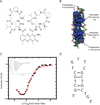
Figure 1. Biochemical analysis of Actinomycin D molecules bound to CTG:CTG DNA
(A) Chemical structure of Actinomycin D. (B) Crystal structure of the 2:1 ActD–(ATGCTGCAT)2 complex (reimaged from Hou et al., 2002). ActD molecules are in blue van der Waals representations, while DNA is shown in skeletal form. Intercalation of the phenoxazone ring is observed at both GpC steps, while pentapeptides remain within the minor groove. (C) Representative ITC isotherm for the interaction between ActD and (CTG)4. Raw heats of reaction versus time are imbedded in the upper left of the graph. The average thermodynamic binding parameters when fit with the sequential binding model are K1 = (9±6)*104, ΔH1 = (7±6)*103 kcal/mol, K2 = (2±1)*106, ΔH2 = (−3±1)*104 kcal/mol. (D) The lowest free energy structure of the (CTG)4 sequence (Reuter et al., 2010).
Figure 2
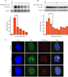
Figure 2. Actinomycin D reduces CUG repeat levels in DM1 cell models
(A-B) CUG repeat RNA levels after ActD treatment in a DM1 HeLa cell system. Northern blot analysis and quantification demonstrate a significant decrease in CUG repeats at 5 nM within the HeLa cells and a dose-dependent decrease follows thereafter. GAPDH, a housekeeping gene, is used as a control. In DM1 patient cells, a modest increase in CUG repeats is observed after 6 nM, and CUG levels persist at that amount from 7 nM to 100 nM ActD (only data points up to 10 nM ActD are shown). Asterisks indicate level of significance (*= p < 0.05, ***= p < 0.001). Error bars represent standard deviation. (C) CUG ribonuclear foci formation in response to ActD treatment in HeLa cells. (top row) Nuclei of untreated HeLa cells. DAPI staining of nucleus is on far left and nuclear MBNL1 staining is in the second column. These untreated cells were not transfected with CUG repeats. (middle row) Transfection with CUG960 gives rise to ribonuclear foci in HeLa cells. Aggregation of MBNL1 and CUG repeats observed in the second and third columns, respectively. (bottom row) Treatment with 10 nM ActD in HeLa cells results in decreased number and size of MBNL1 and CUG aggregates (second and third columns), and reduction of MBNL sequestration as evidence by reversion to more diffuse staining (second column).
Figure 3
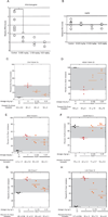
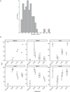
Figure 3. Actinomycin D rescues mis-splicing events in HSALR mice
(A) qRT-PCR analysis of HSA transgene levels in HSALR mice treated with PBS and 0.5% DMSO or indicated dosages of ActD for 5 days. Each circle represents the transcript levels within vastus muscle of a single mouse. Mice treated with any of the ActD dosages exhibited significant reduction of HSA transgene levels as compared to control treated mice. Asterisk indicate significance (* = p < 0.05). (B) qRT-PCR analysis of endogenous DMPK levels (no repeats) in HSALR mice treated with PBS and 0.5% DMSO or indicated dosages of ActD for 5 days. None of the ActD dosages caused significant reduction of DMPK levels. (C-H) Jitterplot representation of various endogenous splicing events perturbed in DM1 mice. Each symbol represents the splicing outcome for vastus muscle of a single mouse, while line represents the average of all experiments. (C) Clcn1 splicing demonstrates almost complete rescue by 0.25 mg kg−1 per day for 5 days. Gel images of two replicates per condition are demonstrated below. (D) In the Atp2a1 event, almost full rescue is observed by 0.25 mg kg−1 per day for 5 days. (E) Mbnl1 auto-regulates exon 5 inclusion, but the highest ActD dosage only reverses ~50% of the observed mis-splicing in mice. (F) ActD reverses mis-splicing of the Vps39 exon 3 event completely by 0.25 mg kg−1 per day for 5 days. (G-H) Nfix and Ldb3 events both exhibit partial but significant reversal of mis-splicing upon treatment with ActD.
Figure 4
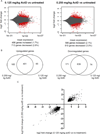
Figure 4. Less than 5% of genes are differentially expressed in mice upon Actinomycin D treatment
(A) MA-plots of 0.125 mg kg−1 compared to no-treatment (PBS) control mice (left panel) and 0.250 mg kg−1 compared to no-treatment (PBS) control mice (right panel). Gene log2 fold change is plotted against the mean of normalized counts. Red circles denote genes with adjusted p values less than 0.1. Points that do not fall within the window are denoted with triangles. (B) Venn diagram depicts the number of differentially expressed genes shared by the two treatments compared to the no-treatment control. (C) Genes are plotted with the log2 fold change for mice treated with low (0.125 mg kg−1) verses high (0.250 mg kg−1) dose of ActD.
Figure 5
Figure 5. Numerous mis-spliced events are rescued upon Actinomycin D treatment in DM1 mice
(A) Distribution of the average percent rescue observed for the 70 splicing events that were mis-regulated in HSALR mice and showed evidence of rescue in littermate mice treated with 0.25 mg kg−1 ActD. (B) Jitterplot representation of various splicing events perturbed in HSALR mice that showed evidence of rescue. Each symbol represents the splicing outcome estimate generated by MISO in the vastus muscle of a single mouse and the line represents the 95% confidence interval on that estimate.

References

    1. Anders S, Pyl PT, Huber W. HTSeq A Python framework to work with high-throughput sequencing data. Bioinformatics. 2015;31:166–169. - PMC - PubMed
    1. Arambula JF, Ramisetty SR, Baranger AM, Zimmerman SC. A simple ligand that selectively targets CUG trinucleotide repeats and inhibits MBNL protein binding. Proc. Natl. Acad. Sci. U.S.A. 2009;106:16068–16073. - PMC - PubMed
    1. Bensaude O. Inhibiting eukaryotic transcription: Which compound to choose? How to evaluate its activity? Transcription. 2011;2:103–108. - PMC - PubMed
    1. Chaouch S, Mouly V, Goyenvalle A, Vulin A, Mamchaoui K, Negroni E, Di Santo J, Butler-Browne G, Torrente Y, Garcia L, et al. Immortalized skin fibroblasts expressing conditional MyoD as a renewable and reliable source of converted human muscle cells to assess therapeutic strategies for muscular dystrophies: validation of an exon-skipping approach to restore dystrophin in Duchenne muscular dystrophy cells. Hum Gene Ther. 2009;20:784–790. - PubMed
    1. Chi LM, Lam SL. Structural roles of CTG repeats in slippage expansion during DNA replication. Nucleic Acids Res. 2005;33(5):1604–1617. - PMC - PubMed

Publication types

MeSH terms

LinkOut - more resources