. 2016 Dec;53(10):7298-7311.
doi: 10.1007/s12035-015-9583-6. Epub 2015 Dec 21.

bFGF Protects Against Blood-Brain Barrier Damage Through Junction Protein Regulation via PI3K-Akt-Rac1 Pathway Following Traumatic Brain Injury

Affiliations

bFGF Protects Against Blood-Brain Barrier Damage Through Junction Protein Regulation via PI3K-Akt-Rac1 Pathway Following Traumatic Brain Injury

Zhou-Guang Wang et al. Mol Neurobiol. 2016 Dec.

Abstract

Many traumatic brain injury (TBI) survivors sustain neurological disability and cognitive impairments due to the lack of defined therapies to reduce TBI-induced blood-brain barrier (BBB) breakdown. Exogenous basic fibroblast growth factor (bFGF) has been shown to have neuroprotective function in brain injury. The present study therefore investigates the beneficial effects of bFGF on the BBB after TBI and the underlying mechanisms. In this study, we demonstrate that bFGF reduces neurofunctional deficits and preserves BBB integrity in a mouse model of TBI. bFGF suppresses RhoA and upregulates tight junction proteins, thereby mitigating BBB breakdown. In vitro, bFGF exerts a protective effect on BBB by upregulating tight junction proteins claudin-5, occludin, zonula occludens-1, p120-catenin, and β-catenin under oxygen glucose deprivation/reoxygenation (OGD) in human brain microvascular endothelial cells (HBMECs). Both the in vivo and in vitro effects are related to the activation of the downstream signaling pathway, PI3K/Akt/Rac-1. Inhibition of the PI3K/Akt or Rac-1 by specific inhibitors LY294002 or si-Rac-1, respectively, partially reduces the protective effect of bFGF on BBB integrity. Overall, our results indicate that the protective role of bFGF on BBB involves the regulation of tight junction proteins and RhoA in the TBI model and OGD-induced HBMECs injury, and that activation of the PI3K/Akt /Rac-1 signaling pathway underlies these effects.

Keywords: Blood-brain barrier; PI3K/Akt /Rac-1; RhoA; Tight junction; Traumatic brain injury; bFGF.


Figures

Fig. 1
Fig. 1
Effects of exogenous bFGF (15 μg) or PBS on Garcia scores (a) at 24 h after TBI. Effects of bFGF on Evans Blue fluorescence (b, c) and FITC-dextran permeability (d) at 24 h after TBI. *P < 0.05 versus the sham group. #P < 0.05 versus the TBI group. Data are the mean values ± SEM, n = 6
Fig. 2
Fig. 2
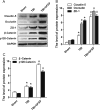
Protein expression of claudin-5, occludin, zonula occludens-1, p120-catenin, and β-catenin for the sham, TBI, and bFGF treatment groups. GAPDH was used as the loading control and for band density normalization (a). The optical density analysis of claudin-5, occludin, zonula occludens-1, p120-catenin and β-catenin protein (b, c). *P < 0.05 versus the sham group. #P < 0.05 versus the TBI group. Data are the mean values ± SEM, n = 6
Fig. 3
Fig. 3
Dual-label immunofluorescence staining results of endothelial cell marker (a) CD31 (red) and different tight junction proteins (b) Claudin-5 (c) Occludin, and (d) ZO-1 in the mouse brain 1 day after TBI. The nuclei are labeled by Hoechst. Scale bar = 10 μm. The proteins with obvious bright signals are labeled. Magnification was ×40
Fig. 4
Fig. 4
Effects of the PI3K/Akt inhibitor LY294002 (50 nmol/kg) on bFGF induced attenuation of brain injury at 24 h after TBI. Evaluation of Garcia test (a) and Evans Blue fluorescence (b, c) and FITC-dextran permeability (d) in mice subjected to TBI. *P < 0.05 versus the sham group. #P < 0.05 versus the TBI group. &P < 0.05 versus the TBI + bFGF group. Data are the mean values ± SEM, n = 6
Fig. 5
Fig. 5
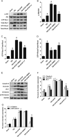
The protein expression of p-Akt, GTP-Rac1, and GTP-RhoA after TBI-induced BBB destruction treated with bFGF and LY294002. GAPDH was used as the loading control and for band density normalization (a). The optical density analysis of p-Akt, GTP-Rac1, and GTP-RhoA protein (b, c). The protein expression of claudin-5, occluding, ZO-1, p120-catenin, and β-catenin after TBI-induced BBB destruction treated with bFGF and LY294002. GAPDH was used as the loading control and for band density normalization (d). The optical density analysis of pclaudin-5, occluding, ZO-1, p120-catenin, and β-catenin protein (e, f). *P < 0.05 versus the sham group. #P < 0.05 versus the TBI group. &P < 0.05 versus the TBI + bFGF group. Data are the mean values ± SEM, n = 6
Fig. 6
Fig. 6
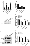
HBMECs were pre-treated with 50 ng/ml bFGF with or without inhibitor LY294002 (20 μM) for 1 h, and then OGD conditions for 24 h and reperfusion for 12 h. The cells were analyzed for FITC-dextran transport (a). The cell lysates were analyzed by western blotting for the expression of CD31, claudin-5, occluding, ZO-1, p120-catenin, and β-catenin, and GAPDH was used as the loading control and for band density normalization (b, c). The optical density analysis of CD31, claudin-5, occluding, ZO-1, p120-catenin and β-catenin protein (c, e, f). *P<0.05 versus the control group. #P<0.05 versus the OGD group. &P<0.05 versus the OGD + bFGF group. Data are the mean values ± SEM, n=6
Fig. 7
Fig. 7
HBMECs were pre-treated with bFGF for 1 h, and then OGD conditions for 24 h, and reperfusion for 12 h. Immunofluorescence staining of confluent HBMECs monolayers for (a) claudin-5, (b) occluding, (c) p120-catenin, and (d) β-catenin. Nuclei were labeled by Hoechst. The proteins with obvious bright signals are labeled. Magnification was ×40
Fig. 8
Fig. 8
The protein expression of p-Akt, GTP-Rac1 and GTP-RhoA in OGD-induced HBMECs treated with bFGF and LY294002 inhibitor. GAPDH was used as the loading control and for band density normalization (a). The optical density analysis of p-Akt, GTP-Rac1 and GTP-RhoA protein (b, c, d). *P < 0.05 versus the control group. #P < 0.05 versus the OGD group. &P < 0.05 versus the OGD + bFGF group. Data are the mean values ± SEM, n = 6
Fig. 9
Fig. 9
The protein expression of claudin-5, occluding, ZO-1, p120-catenin, β-catenin, GTP-Rac1 and GTP-RhoA in HBMECs was determined by Western blotting. GAPDH was used as the loading control and for band density normalization (a). The optical density analysis of all these proteins (b, c, d, e). *P < 0.05 versus the corresponds group. #P < 0.05 versus the control group. &P < 0.05 versus the OGD group. Data are the mean values ± SEM, n = 6

References

    1. Maas AI, Stocchetti N, Bullock R. Moderate and severe traumatic brain injury in adults. Lancet Neurol. 2008;7(8):728–741. doi: 10.1016/S1474-4422(08)70164-9. - DOI - PubMed
    1. Coronado VG, Xu L, Basavaraju SV, McGuire LC, Wald MM, Faul MD, Guzman BR, Hemphill JD. Surveillance for traumatic brain injury-related deaths—United States, 1997-2007. MMWR Surveill Summ. 2011;60(5):1–32. - PubMed
    1. Shlosberg D, Benifla M, Kaufer D, Friedman A. Blood-brain barrier breakdown as a therapeutic target in traumatic brain injury. Nat Rev Neurol. 2010;6(7):393–403. doi: 10.1038/nrneurol.2010.74. - DOI - PMC - PubMed
    1. Nag S, Kapadia A, Stewart DJ. Review: molecular pathogenesis of blood-brain barrier breakdown in acute brain injury. Neuropathol Appl Neurobiol. 2011;37(1):3–23. doi: 10.1111/j.1365-2990.2010.01138.x. - DOI - PubMed
    1. Werner C, Engelhard K. Pathophysiology of traumatic brain injury. Br J Anaesth. 2007;99(1):4–9. doi: 10.1093/bja/aem131. - DOI - PubMed

MeSH terms