. 2016 Jun;33(3):447-56.
doi: 10.1007/s10719-015-9638-y. Epub 2015 Dec 19.

Glycoproteomic studies of IgE from a novel hyper IgE syndrome linked to PGM3 mutation

Affiliations

Glycoproteomic studies of IgE from a novel hyper IgE syndrome linked to PGM3 mutation

Gang Wu et al. Glycoconj J. 2016 Jun.

Abstract

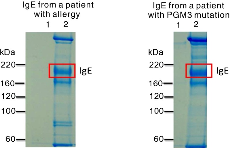
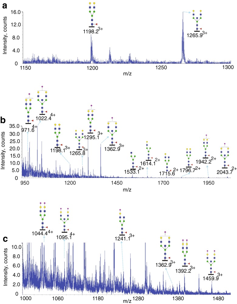
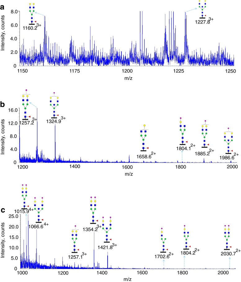
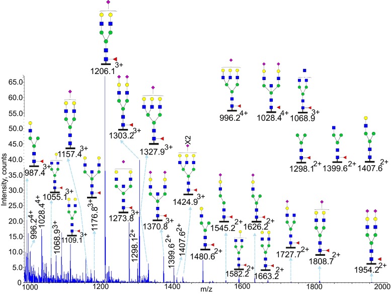
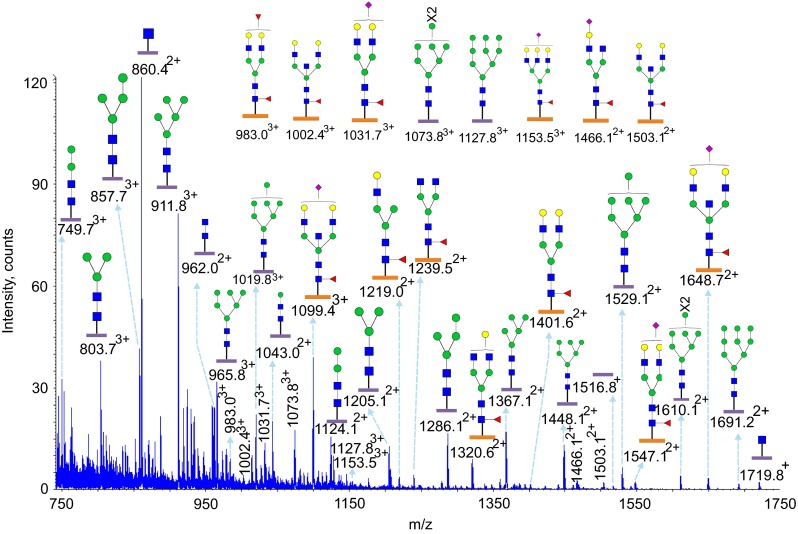
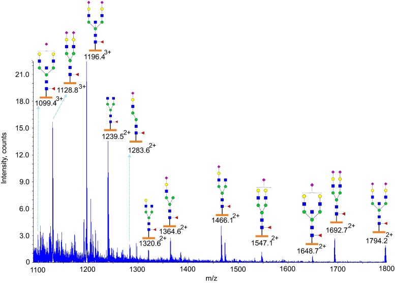
Glycans serve as important regulators of antibody activities and half-lives. IgE is the most heavily glycosylated antibody, but in comparison to other antibodies little is known about its glycan structure function relationships. We therefore describe the site specific IgE glycosylation from a patient with a novel hyper IgE syndrome linked to mutations in PGM3, which is an enzyme involved in synthesizing UDP-GlcNAc, a sugar donor widely required for glycosylation. A two-step method was developed to prepare two IgE samples from less than 1 mL of serum collected from a patient with PGM3 mutation and a patient with atopic dermatitis as a control subject. Then, a glycoproteomic strategy was used to study the site-specific glycosylation. No glycosylation was found at Asn264, whilst high mannose glycans were only detected at Asn275, tri-antennary glycans were exclusively observed at Asn99 and Asn252, and non-fucosylated complex glycans were detected at Asn99. The results showed similar glycosylation profiles between the two IgE samples. These observations, together with previous knowledge of IgE glycosylation, imply that IgE glycosylation is similarly regulated among healthy control, allergy and PGM3 related hyper IgE syndrome.

Keywords: Glycoproteomics; Hyper IgE Syndrome; IgE; Phosphoglucomutase 3.


Figures

Fig. 1
Fig. 1
SDS-PAGE of immunoprecipitated IgE. 600 ul of serum from a patient with atopic dermatitis as a control subject and 600 ul of plasma from a patient with hyper IgE syndrome were used to prepare IgE. Elutions of immunoprecipitation were analyzed by non-reduced SDS-PAGE in 3–8 % Tris-acetate gel with Tris-acetate running buffer. Lane 1: Control resin. Lane 2: Immunoprecipitated IgE. A band was observed between 220 and 160 kDa in Lane 2, which is consistent with the molecular weight of IgE
Fig. 2
Fig. 2
Glycosylation of IgE Asn21 from a PGM3 patient. Data were acquired by LC/MS. The glycopeptides were produced by chymotrypsin digestion. The peptide backbone is presented as a black bar under each glycan (12TRCCKNIPSNATSVTL27). The ions are in the form of M+nHn+. Glycopeptides were detected eluting at three time points: from 30.5 to 31.5 min (a), from 32.5 to 33.5 min (b) and from 35 to 37 min (c). The glycan structures were deduced according to the molecular weight, fragmention of glycopeptides by MS/MS analysis, previous glycomic analysis of released IgE N-glycans and knowledge of N-glycosylation biosynthetic pathways
Fig. 3
Fig. 3
Glycosylation of IgE Asn49 from a PGM3 patient. Data were acquired by LC/MS. The glycopeptides were produced by chymotrypsin digestion. The peptide backbone (44DTGSLNGTTMTLPATTL60) is presented as a black bar under each glycan. The ions are in the form of M+nHn+. Glycopeptides were detected eluting at three time points: from 37 to 38 min (a), from 40 to 42 min (b) and from 46 to 47.5 min (c). The glycan structures were deduced according to the molecular weight, fragmention of glycopeptides by MS/MS analysis, previous glycomic analysis of released IgE N-glycans and knowledge of N-glycosylation biosynthetic pathways
Fig. 4
Fig. 4
Glycosylation of IgE Asn99 from a PGM3 patient. Data were acquired by LC/MS. The glycopeptides were produced by trypsin digestion. The peptide backbone (87VAHTPSSTDWVDNK100) is presented as a black bar under each glycan. The ions are in the form of M+nHn+. Glycopeptides were detected eluting between 21 and 23 min. The glycan structures were deduced according to the molecular weight, fragmention of glycopeptides by MS/MS analysis, previous glycomic analysis of released IgE N-glycans and knowledge of N-glycosylation biosynthetic pathways
Fig. 5
Fig. 5
Glycosylation of IgE Asn252 and Asn275 from a PGM3 patient. Data were acquired by LC/MS. The glycopeptides were produced by trypsin digestion. Peptide backbones with Asn252 (249GTVNLTWSR257) and Asn275 (275 NGTLTVTSTLPVGTR289) are presented as an orange bar and a purple bar respectively under each glycan. The ions are in the form of M+nHn+. Glycopeptides were eluted between 27.5 and 32 min. The glycan structures were deduced according to the molecular weight, fragmention of glycopeptides by MS/MS analysis, previous glycomic analysis of released IgE N-glycans and knowledge of N-glycosylation biosynthetic pathways
Fig. 6
Fig. 6
Glycosylation of IgE Asn252 from a PGM3 patient. Data were acquired by LC/MS. The glycopeptides were produced by trypsin digestion. The peptide backbone with (249GTVNLTWSR257) is presented as an orange bar under each glycan. The ions are in the form of M+nHn+. The glycopeptides were eluted from 34.5 to 36.5 min. The glycan structures were deduced according to the molecular weight, fragmention of glycopeptides by MS/MS analysis, previous glycomic analysis of released IgE N-glycans and knowledge of N-glycosylation biosynthetic pathways

References

    1. Arnold JN, Wormald MR, Sim RB, Rudd PM, Dwek RA. The impact of glycosylation on the biological function and structure of human immunoglobulins. Annu. Rev. Immunol. 2007;25:21–50. doi: 10.1146/annurev.immunol.25.022106.141702. - DOI - PubMed
    1. Parekh RB, Dwek RA, Sutton BJ, Fernandes DL, Leung A, Stanworth D, Rademacher TW, Mizuochi T, Taniguchi T, Matsuta K, et al. Association of rheumatoid arthritis and primary osteoarthritis with changes in the glycosylation pattern of total serum IgG. Nature. 1985;316(6027):452–457. doi: 10.1038/316452a0. - DOI - PubMed
    1. Anthony RM, Wermeling F, Ravetch JV. Novel roles for the IgG Fc glycan. Ann. N. Y. Acad. Sci. 2012;1253:170–180. doi: 10.1111/j.1749-6632.2011.06305.x. - DOI - PubMed
    1. Scallon BJ, Tam SH, McCarthy SG, Cal AN, Raju TS. Higher levels of sialylated Fc glycans in immunoglobulin G molecules can adversely impact functionality. Mol. Immunol. 2007;44(7):1524–1534. doi: 10.1016/j.molimm.2006.09.005. - DOI - PubMed
    1. Anthony RM, Wermeling F, Karlsson MC, Ravetch JV. Identification of a receptor required for the anti-inflammatory activity of IVIG. Proc. Natl. Acad. Sci. U. S. A. 2008;105(50):19571–19578. doi: 10.1073/pnas.0810163105. - DOI - PMC - PubMed

Publication types

MeSH terms