. 2015 Dec 17;163(7):1702-15.
doi: 10.1016/j.cell.2015.11.056.

Immunogenicity of Stabilized HIV-1 Envelope Trimers with Reduced Exposure of Non-neutralizing Epitopes

Affiliations

Immunogenicity of Stabilized HIV-1 Envelope Trimers with Reduced Exposure of Non-neutralizing Epitopes

Steven W de Taeye et al. Cell. .

Abstract

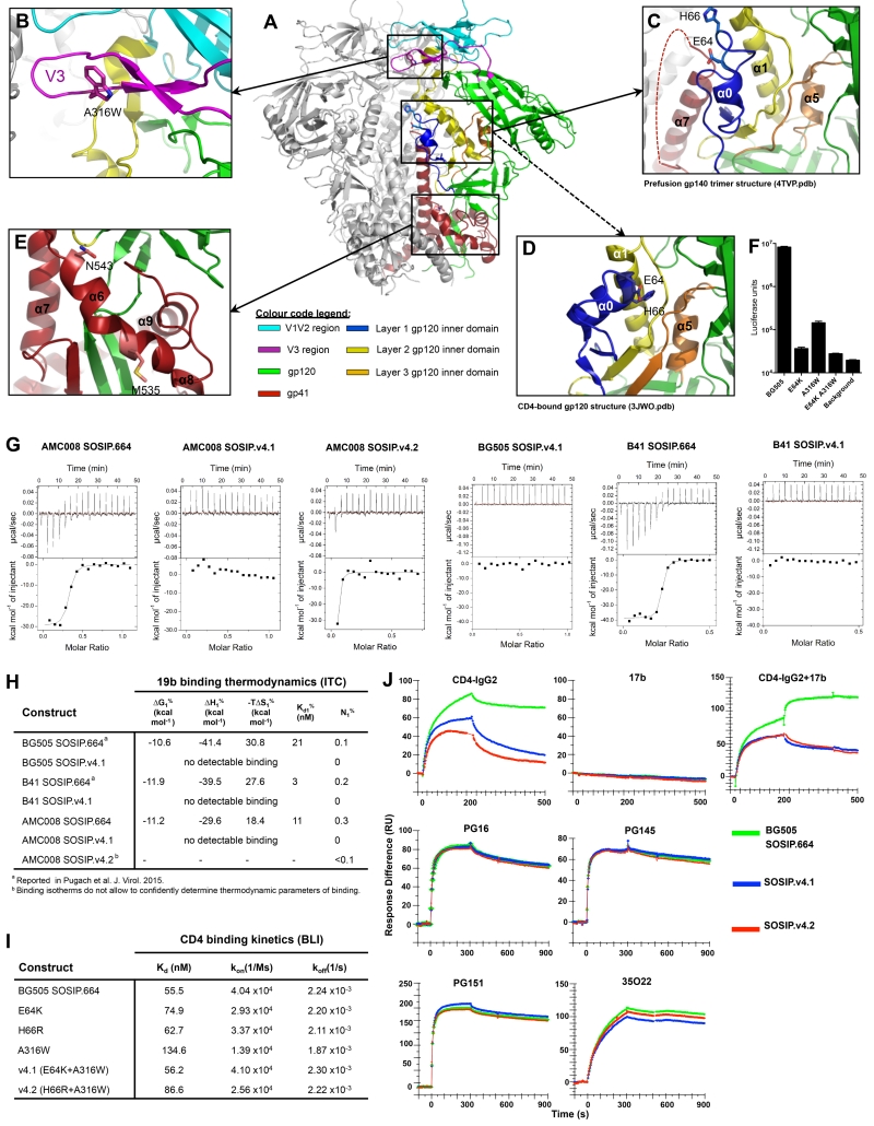
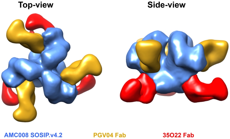
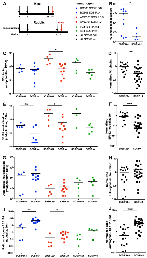
The envelope glycoprotein trimer mediates HIV-1 entry into cells. The trimer is flexible, fluctuating between closed and more open conformations and sometimes sampling the fully open, CD4-bound form. We hypothesized that conformational flexibility and transient exposure of non-neutralizing, immunodominant epitopes could hinder the induction of broadly neutralizing antibodies (bNAbs). We therefore modified soluble Env trimers to stabilize their closed, ground states. The trimer variants were indeed stabilized in the closed conformation, with a reduced ability to undergo receptor-induced conformational changes and a decreased exposure of non-neutralizing V3-directed antibody epitopes. In rabbits, the stabilized trimers induced similar autologous Tier-1B or Tier-2 NAb titers to those elicited by the corresponding wild-type trimers but lower levels of V3-directed Tier-1A NAbs. Stabilized, closed trimers might therefore be useful components of vaccines aimed at inducing bNAbs.


Figures

Figure 1
Figure 1. Design of amino-acid substitutions to stabilize SOSIP.664 trimers
(A) Crystal structure of BG505 SOSIP.664 trimer (Pancera et al., 2014), showing the locations of amino-acid substitutions relevant to this study. One protomer is colored according to sub-regions: gp41 in red; V1V2 in cyan; V3 in purple; gp120 inner domain layer 1 in blue; layer 2 in yellow; layer 3 in orange; outer domain and N- and C- termini of gp120 in green. (B) Detailed view of V3 and surrounding regions showing the A316W substitution. (C) Detailed view of layers 1 and 2 of the gp120 inner domain, showing how E64 and H66 face the unstructured region of gp41 (residues 548-568) between α6 and α7. (D) Detailed view of a CD44-liganded gp120 monomer structure (Pancera et al., 2010) highlighting the same region as in (C) and showing how the E64 and H66 side chains face gp120 layer 2. (E) Depiction of how the α6-α9 helices of gp41 encircle the N- and C-termini of gp120, with M535 and N543 located in the middle and at the C-terminus of α6, respectively. (F) Infection of TZM-bl cells by BG505.T332N Env-pseudoviruses with an A316W, E64K or H66R substitution. (G) V3 Fab 19b binding to AMC008, BG505 and B41 SOSIP.664 and SOSIP.v4 trimers was assessed by ITC. The enthalpy changes (ΔH), dissociation constants (KD) and stoichiometries of binding (molar ratio; N) are listed in panel (H). (I) Biolayer interferometry analysis of CD4 binding to AviB-tagged mutant wild-type BG505 SOSIP.664 trimers. (J) SPR analysis of ligand binding to BG505 SOSIP.664-His trimers and both versions of SOSIP.v4-His trimers. Top row: CD4- IgG2 (left panel), 17b (middle panel), CD4-IgG2 followed by 17b (right panel); bottom row, PG16, PGT145, PGT151 and 35O22. Antibodies and CD4-IgG2 were injected at 500 nM. See also figure S1, table S2 and S3.
Figure 2
Figure 2. HDX-MS analysis of BG505 SOSIP.664 and SOSIP.v4 trimers
Butterfly plots of PGT145-purified BG505 (A) SOSIP.664, (B) SOSIP.v4.1, (C) SOSIP.v4.2 trimers, comparing deuterium exchange levels in the presence and absence of sCD4. The percent exchange at each time point for each peptide is shown at its position along the primary sequence of the protein. Regions that are less (red) or more (blue) protected upon CD4-binding are mapped on the BG505 SOSIP.664 crystal structure (Pancera et al., 2014), to depict CD4-induced conformational changes. The differences in CD4-induction between the SOSIP.664 and SOSIP.v4.1 or SOSIP.v4.2 trimers were also plotted on the crystal structure (rightmost panels of B and C). The exchange kinetics of individual peptides are shown in Fig. S4 and Data S1.
Figure 3
Figure 3. EM reconstruction of AMC008 SOSIP.v4.2 trimers in complex with PGV04 and 35O22 Fabs
Top and side view of AMC008.v4.2 trimers (blue) in complex with PGV04 (yellow) and 35O22 (red) Fabs. The reconstruction shows that AMC008.v4.2 trimers are compact and native-like. At the resolution obtained here, there is a very high degree of structural similarity with BG505 SOSIP.664 trimers in complex with 35O22 [EMDB-2672). See also figure S5.
Figure 4
Figure 4. Immunogenicity of AMC008, BG505 and B41 SOSIP.v4 trimers
(A) Immunization schedules. Mice were immunized at weeks 0, 4 and 16 (black arrows) and the Ab responses were analyzed at week 18 (red arrow). Rabbits were immunized at weeks 0, 4 and 20 (black arrows) and the Ab responses were analyzed at week 22 (red arrow). Blue symbols represent BG505 trimer-immunized animals, red symbols AMC008 and green symbols B41. The SOSIP.664 recipients are shown by closed circles, SOSIP.v4 by squares. Statistical comparisons between groups were performed using a two-tailed Mann-Whitney U test (*p>0.05, **p>0.01, ***p>0.001). (B) V3 peptide ELISA endpoint binding Ab titers in mouse sera are plotted. (C, D) V3 peptide binding responses were determined by ELISA. The midpoint binding Ab titers in rabbit sera are plotted in (C), and the normalized values of all SOSIP.664 and SOSIP.v4 values, compared to the mean of the corresponding SOSIP.664 group, are shown in (D). The midpoint (E, F) SF162 (Tier-1A), and (G, H) autologous Tier-2 (BG505 and B41) or Tier-1B (AMC008) neutralization titers were determined using the TZM-bl assay (Tables S4 and S5). Specifically, the serum dilutions at which HIV-1 infectivity is inhibited by 50% (ID50) are plotted in (E) and (G), and the normalized values of all SOSIP.664 and SOSIP.v4 values, compared to the mean of the corresponding SOSIP.664 group, are shown in (F) and (H). (I) The ratios of autologous/SF162 NAb titers are shown for sera from the various SOSIP.664 and SOSIP.v4 immunized rabbits. (J) The normalized values derived from the data in (I) are plotted. See also figure S6 and table S4 and S5.

References

    1. Binley JM, Sanders RW, Clas B, Schuelke N, Master A, Guo Y, Kajumo F, Anselma DJ, Maddon PJ, Olson WC, et al. A recombinant human immunodeficiency virus type 1 envelope glycoprotein complex stabilized by an intermolecular disulfide bond between the gp120 and gp41 subunits is an antigenic mimic of the trimeric virion-associated structure. J. Virol. 2000;74:627–643. - PMC - PubMed
    1. Binley JM, Sanders RW, Master A, Cayanan CS, Wiley CL, Schiffner L, Travis B, Kuhmann S, Burton DR, Hu S, et al. Enhancing the proteolytic maturation of human immunodeficiency virus type 1 envelope glycoproteins. J. Virol. 2002;76:2606–2616. - PMC - PubMed
    1. Blattner C, Lee J, Sliepen K, Derking R, Falkowska E, DelaPeña A, Cupo A, Julien JP, VanGils M, Lee PS, et al. Structural delineation of a quaternary, cleavage-dependent epitope at the gp41-gp120 interface on intact HIV-1 env trimers. Immunity. 2014;40:669–680. - PMC - PubMed
    1. Bonomelli C, Doores KJ, Dunlop DC, Thaney V, Dwek RA, Burton DR, Crispin M, Scanlan CN. The glycan shield of HIV is predominantly oligomannose independently of production system or viral clade. PLoS One. 2011;6:1–7. - PMC - PubMed
    1. Chaudhury S, Reifman J, Wallqvist A. Simulation of B cell affinity maturation explains enhanced antibody cross-reactivity induced by the polyvalent malaria vaccine AMA1. J. Immunol. 2014;193:2073–2086. - PMC - PubMed

Publication types