. 2015 Dec 17;163(7):1783-1795.
doi: 10.1016/j.cell.2015.11.060.

Genetic Identification of an Expansive Mechanoreceptor Sensitive to Skin Stroking

Affiliations

Genetic Identification of an Expansive Mechanoreceptor Sensitive to Skin Stroking

Ling Bai et al. Cell. .

Abstract

Touch perception begins with activation of low-threshold mechanoreceptors (LTMRs) in the periphery. LTMR terminals exhibit tremendous morphological heterogeneity that specifies their mechanical receptivity. In a survey of mammalian skin, we found a preponderance of neurofilament-heavy-chain(+) circumferential endings associated with hair follicles, prompting us to develop a genetic strategy to interrogate these neurons. Targeted in vivo recordings revealed them to be Aβ field-LTMRs, identified 50 years ago but largely elusive thereafter. Remarkably, while Aβ field-LTMRs are highly sensitive to gentle stroking of the skin, they are unresponsive to hair deflection, and they encode skin indentation in the noxious range across large, spotty receptive fields. Individual Aβ field-LTMRs form up to 180 circumferential endings, making them the most anatomically expansive LTMR identified to date. Thus, Aβ field-LTMRs are a major mammalian LTMR subtype that forms circumferential endings in hairy skin, and their sensitivity to gentle skin stroking arises through integration across many low-sensitivity circumferential endings.


Figures

Figure 1
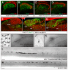
Figure 1. Genetic labeling of sensory neurons that innervate the dorsal column nuclei
(A-D) The C1 DC of R26LSL-tdTomato mice was injected with AAV2/1-Cre virus to retrogradely label neurons that project to the DCN. (B-D) In hairy skin, at least 4 types of terminals are labeled by tdTomato: an unknown ending type innervating S100+ terminal organs that resemble Pacinian corpuscles (B), Merkel endings from Aβ SA1-LTMRs that associate with Troma1+ Merkel cells (C), lanceolate endings that are NFH+ (97.8 ± 1.2%, 156 terminals, n = 3) and derived from Aβ RA-LTMRs (D, asterisk), as well as circumferential endings that are also NFH+ (99.5% ± 0.5%, 465 terminals, n = 3) (D, arrowhead). (E) NFH+ circumferential endings, NFH+ lanceolate endings, and Merkel endings innervate 94 ± 3%, 8 ± 2%, and 0.8 ± 0.2% of hair follicles, respectively (3964 hair follicles, n = 3). (F-I) Immunostaining of NFH and Tuj1 on hairy skin sections reveals that NFH+ circumferential endings (arrowhead) can be found in mouse (F), cat (G), dog (H), and macaue(I). (J) Diagram of the TrkC and Ret intersectional genetic labeling strategy. (K) Double immunostaining of hairy skin sections from TrkCCreER; Retf(CFP) mice treated with 0.1 tamoxifen at P5 reveals that CFP specifically labels the majority of NFH+ circumferential endings (79.6 ± 3.3%, 1142 terminals, n = 4) and all of CFP+ circumferential endings are NFH+ (99.2 ± 0.2%, 904 terminals, n = 4). (L) Whole-mount immunostaining of hairy skin from TrkCCreER; Retf(CFP) mice treated with 3mg tamoxifen at E12.5 reveals that CFP specifically labels Aβ SA1-LTMRs innervating Troma1+ Merkel cells (40 ± 4%, 78 terminals, n = 3). (M) Quantification of the percentage of sensory endings labeled by CFP in K-L. Scale bar: 50 μm (B-D, F-I, K-L). See also Figure S1, S2, and S3.
Figure 2
Figure 2. Neurons with NFH+ circumferential endings innervate the deep spinal cord dorsal horn as well as the DCN
(A-D) Spinal cord sections from TrkCCreER; Retf(CFP) mice treated with 0.1 mg tamoxifen at P5 or 3mg tamoxifen at E12.5. The central projections of neurons with NFH+ circumferential endings labeled by CFP are located in lamina IIiv through IV (n = 9), ventral to lamina IIid labeled by IB4 (A) but partially overlapping with lamina IIiv labeled by PKCγ (B). The central projections of Aβ SA1-LTMRs labeled by CFP are located in lamina III through V (n = 4), ventral to lamina IIid and lamina IIiv labeled by IB4 (C) and PKCγ (D). (E-G) Transverse brainstem sections from TrkCCreER; Retf(CFP) mice treated with 0.1mg tamoxifen at P5 reveals the innervation pattern in the DCN, which is marked by vGlut1. (H-M) Whole-mount AP staining of the skin (H and J), brainstem (I and K), and spinal cord (L and M) from TrkCCreER; Brn3af(AP) mice treated with 0.001mg tamoxifen at P8 reveals the peripheral terminals and central projections of neurons with circumferential endings (H, I and L) or Merkel endings (J, K, and M). Dorsal view of the left DCN and left thoracic spinal cord reveals that axons from a single labeled circumferential ending neuron and Aβ SA1-LTMR project into the spinal cord and form collaterals in both the rostral and caudal directions (L and M). The rostral end of this axon extends to the DCN, where it forms collaterals (I and K). Scale bar: 100 μm (A-G) and 200 μm (H-M). See also Figure S4.
Figure 3
Figure 3. Neurons with NFH+ circumferential endings are Aβ Field-LTMRs
(A) The targeted in vivo DRG recording preparation. (B) Representative responses of Aβ SA-LTMR, Aβ RA-LTMR, and NFH+ circumferential ending neurons to skin stroke delivered by a force controlled brush translated by an ultrasonic piezoelectric stage. The recorded holding force (black, ~5 mN) is shown at top. The stroke speed is 10 mm/sec. (C) Quantification of stroke-evoked action potential firing rates in Aβ SA1-LTMR (n = 4), Aβ RA-LTMR (n = 5), and NFH+ circumferential neurons (n = 10). Responses were computed as the average of the three highest firing rates observed in a 100 msec window which corresponds to the approximate time required for the brush to transit the full arbor of an Aβ-LTMR. NFH+ circumferential ending neurons responded to innocuous stroke with firing rates indistinguishable from those of other hairy skin LTMRs (p = 0.27 Welch’s t-test). (D) The conduction velocities of NFH+ circumferential ending neurons are indistinguishable from those of other hairy skin Aβ RA- and SA-LTMRs recorded (p = 0.871, Welch’s t-test). (E) Von Frey threshold measurements reveal that the force thresholds of NFH+ circumferential ending neurons are higher than those of other Aβ subtypes (90% bootstrap CI [21.2, 52.3] fold higher). (F) Temporal patterns of responses to indentation. Representative peristimulus time histograms (PSTHs) show that NFH+ circumferential neurons adapt more rapidly than Aβ SA1-LTMRs but lack the pronounced off-step responses of the Aβ RA-LTMRs. The indentation force recording (top) is aligned to the PSTH showing the mean firing rate computed across 15 stimulus presentations (7 msec bin widths). (G) Representative spiking responses of NFH+ circumferential ending neurons and Aβ RA-LTMR to deflection of guard hairs. (H) Group data showing that all Aβ RA-LTMRs respond similarly to deflection of guard hairs and skin stroke (p = 0.85, paired t-test), while NFH+ circumferential ending neurons are insensitive to guard hair deflection (p = 0.017, paired t-test). As in (C), evoked firing rates are computed over a 100 msec window. See also Figure S5.
Figure 4
Figure 4. Aβ Field-LTMRs are insensitive to hair deflection yet sensitive to direct stimulation of skin
(A) Laser Doppler Vibrometric measurements of skin and hair movement in response to air puff (left, 15m/sec at the source) and indentation (right, 2g von Frey filament). Shown are representative displacements of the base of a hair shaft and a nearby patch of skin. (B) Group data showing the power spectral density averaged across stimulus presentations and measurement locations (n = 5 hairs, n = 5 skin locations). For air puff, the motion recorded at the base of hairs exceeds that of the surrounding skin by three orders of magnitude. (C) Representative in vivo recordings of Aβ Field-LTMR (blue) and Aβ RA-LTMR (black) responses to air puff and stroke, before and after hair removal. The recording was maintained continuously in all cases. (D) Air puff evoked spiking responses from Aβ Field-LTMRs (n = 6), Aβ SA1-LTMR (n = 3), Aβ RA-LTMRs (n = 4) and Aβ RA-LTMR after hair removal (n = 4). Firing rates computed over the entire 100 msec air puff are plotted against the speed of the air at the stimulator nozzle, which was placed 12mm above the skin. All Aβ RA-LTMR retained sensitivity to stroke after hair removal (C, data not shown), demonstrating that they remained mechanically sensitive despite loss of air puff responses. Traces are offset for clarity.
Figure 5
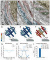
Figure 5. Ultrastructural features of lanceolate and circumferential endings that support the distinct response properties of Aβ RA-LTMRs and Aβ Field-LTMRs
(A-C) TEM images of cross-sections through a lanceolate and circumferential ending complex at a hair follicle. (B-C) Magnified view of the region in the red box of (A). The lanceolate ending (white asterisks) and terminal Schwann cell complexes are embedded in longitudinal oriented collagen fibers (L.C.) in close proximity to hair follicle epithelial cells. Circumferential endings (arrowheads) are embedded in circumferentially oriented collagen fibers (C.C.) in the outer layer. The lanceolate endings, circumferential endings, terminal Schwann cells, and hair follicle epithelia cells are pseudo colored in green, blue, and pink, and yellow, respectively. (D-I) The FEM simulations of the strain acting on lanceolate or circumferential endings in response to mechanical stimuli. (D-F) Top: Contour plots of strain distribution along the lanceolate and circumferential endings in response to 100 μm hair deflection (D), 0.8 mN skin indentation (E), and 10 mN skin indentation (F). Bottom: schematic diagram of the stimulations. (G-H) Plot of the average strain along different positions of lanceolate or circumferential terminals. The zero location is marked by the red asterisk in Figure 6D. (I) The ratio of accumulated strain in the absence and the presence of collagen layers along lanceolate or circumferential endings under either 100 μm hair deflections or 0.8mN skin indentations. Scale bar: 1 μm (A-C). See also Supplemental Table 1.
Figure 6
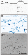
Figure 6. Aβ Field-LTMRs have large receptive fields comprising many weak mechanosensitive endings
(A) In vivo loose patch electrophysiological recordings from an Aβ Field-LTMR in response to 1 mN (left) and 27 mN (right) spatially patterned skin indentations with a 20 μm tipped tungsten probe. The indenter was positioned over the skin in 50 μm steps, such that adjacent steps correspond to adjacent rows in the raster plot. (B) Receptive field of an Aβ Field-LTMR mapped with a 27 mN indentation. Grid indentations of Aβ Field-LTMRs reveal punctuated “hotspots” that extend for 50-100 μm and are separated by insensitive patches (n = 2). (C) Whole-mount AP staining of hairy skin reveals peripheral terminals from individual Aβ Field-LTMRs. Scale bar: 500 μm (B-C). See also Figure S6.
Figure 7
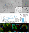
Figure 7. Aβ Field-LTMRs have large terminal fields and a long distance between their axon terminals and SISs
(A-E) Whole-mount AP staining of hairy skin reveals peripheral terminals from individual LTMR subtypes sparsely labeled by a Brn3af(AP) reporter line. (F) Quantification of the number of innervated hair follicles and terminal field area from different LTMR subtypes including Aβ Field-LTMRs (n = 125, 8 mice), Aβ RA-LTMRs (n = 127, 10 mice), Aβ SA1-LTMRs (n = 31, 16 mice), Aδ-LTMRs (n = 55, 4 mice), as well as C-LTMRs (n = 41, 3 mice). Each dot represents a single neuron. (G-I) Whole-mount immunostaining of hairy skin reveals the relationship between MBP, the SIS as inferred from βIV-Spectrin and the terminals of three Aβ-LTMRs subtypes. Arrows or arrowheads point out the SISs or nodes of Ranvier, respectively. Note that the red βIV-Spectrin puncta in G (arrowhead) is associated with a different myelinated axon that is not belong to the genetically labeled Aβ Field-LTMRs. (J) Quantification of the distance of non-myelinated axons reveals that Aβ Field-LTMRs have longer non-myelinated axons (153 ± 50, n = 5) compared to Aβ RA-LTMRs (6.0 ± 1.8, n = 12) (p = 0.03, student t-test) and Aβ SA1-LTMRs (18.8 ± 0.7, n = 8) (p = 0.04, student t-test). Scale bar: 500 μm (A-E). 20 μm (G-I). See also Figure S6 and S7.

References

    1. Abraira VE, Ginty DD. The sensory neurons of touch. Neuron. 2013;79:618–639. - PMC - PubMed
    1. Albers KM, Perrone TN, Goodness TP, Jones ME, Green MA. Cutaneous Overexpression of NT-3 Increases Sensory and Sympathetic Neuron Number and Enhances Touch Dome and Hair Follicle Innervation. J. Cell Biol. 1996;134:487–497. - PMC - PubMed
    1. Badea TC, Cahill H, Ecker J, Hattar S, Nathans J. Distinct roles of transcription factors brn3a and brn3b in controlling the development, morphology, and function of retinal ganglion cells. Neuron. 2009;61:852–864. - PMC - PubMed
    1. Badea TC, Williams J, Smallwood P, Shi M, Motajo O, Nathans J. Combinatorial expression of Brn3 transcription factors in somatosensory neurons: genetic and morphologic analysis. J. Neurosci. 2012;32:995–1007. - PMC - PubMed
    1. Ballermann M, McKenna J, Whishaw IQ. A grasp-related deficit in tactile discrimination following dorsal column lesion in the rat. Brain Res. Bull. 2001;54:237–242. - PubMed

Publication types

LinkOut - more resources