. 2015 Dec 21:5:18519.
doi: 10.1038/srep18519.

Thrombopoietin induces production of nucleated thrombocytes from liver cells in Xenopus laevis

Affiliations

Thrombopoietin induces production of nucleated thrombocytes from liver cells in Xenopus laevis

Yuta Tanizaki et al. Sci Rep. .

Abstract

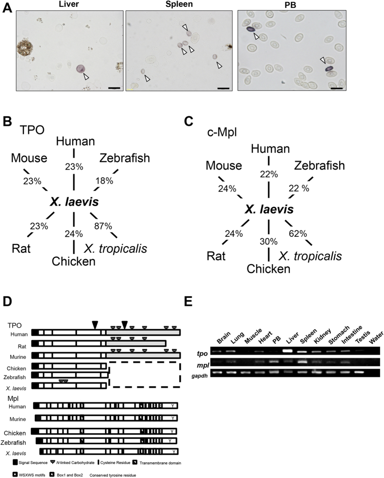
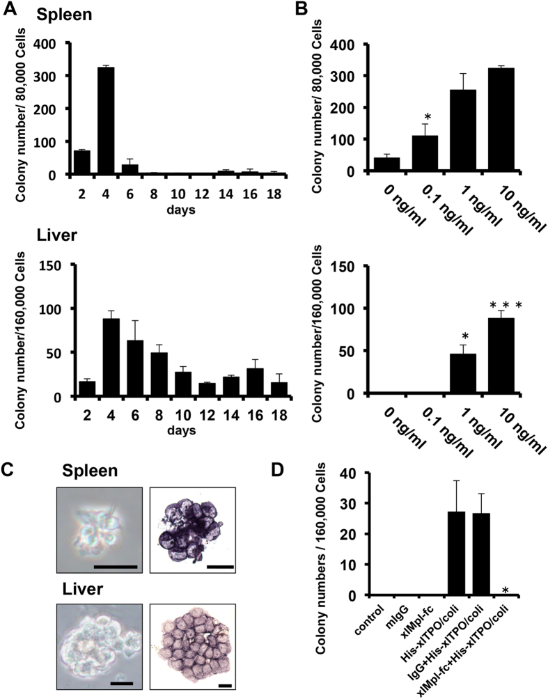
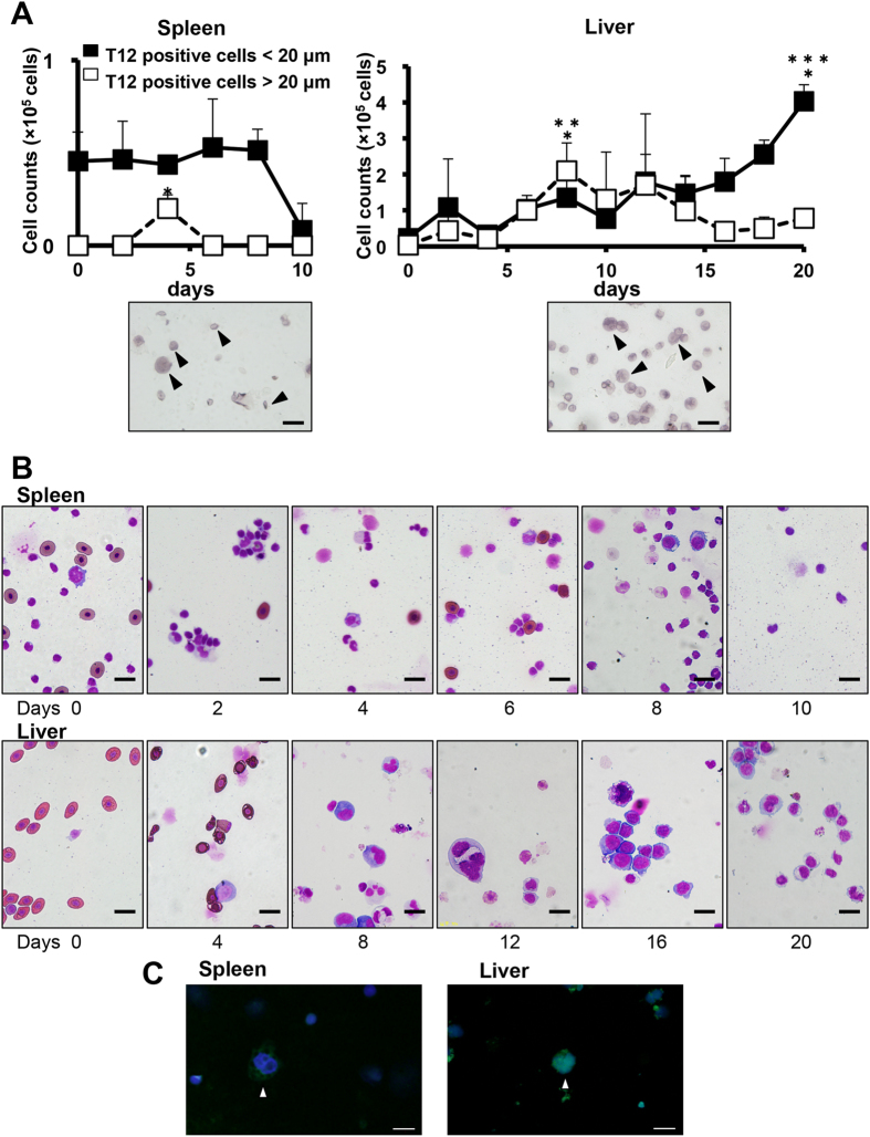
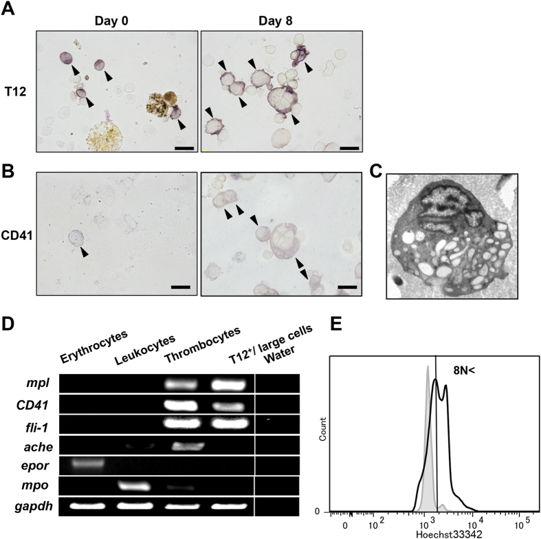
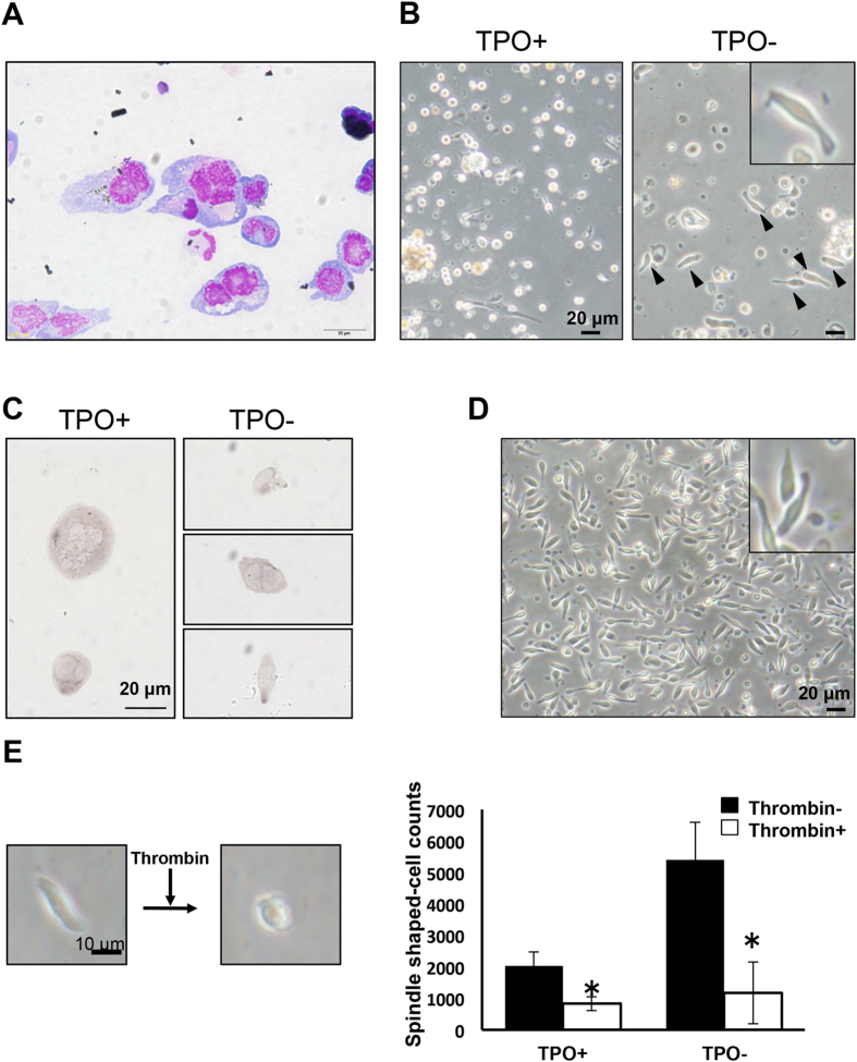
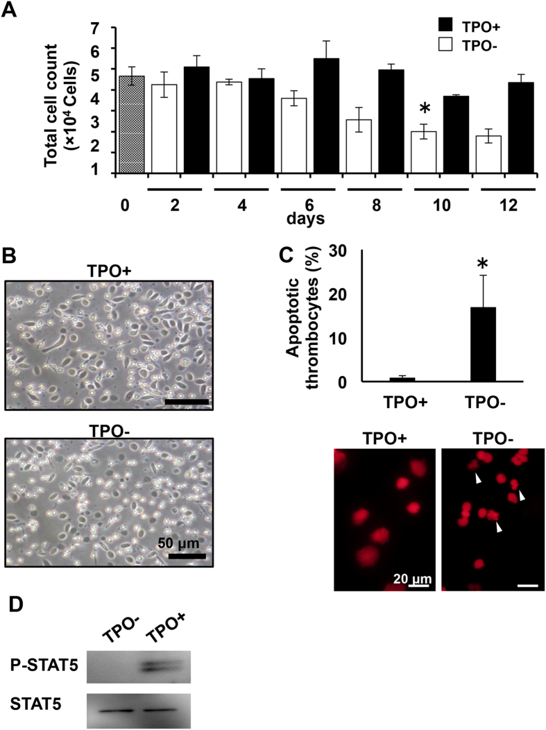
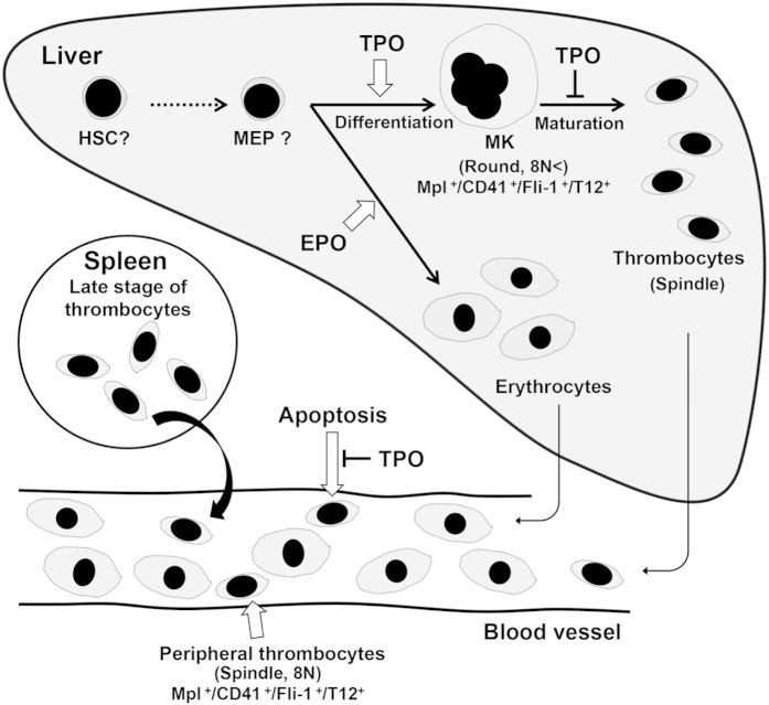
The development of mammalian megakaryocytes (MKs) and platelets, which are thought to be absent in non-mammals, is primarily regulated by the thrombopoietin (TPO)/Mpl system. Although non-mammals possess nucleated thrombocytes instead of platelets, the features of nucleated thrombocyte progenitors remain to be clarified. Here, we provide the general features of TPO using Xenopus laevis TPO (xlTPO). Hepatic and splenic cells were cultured in liquid suspension with recombinant xlTPO. These cells differentiated into large, round, polyploid CD41-expressing cells and were classified as X. laevis MKs, comparable to mammalian MKs. The subsequent culture of MKs after removal of xlTPO produced mature, spindle-shaped thrombocytes that were activated by thrombin, thereby altering their morphology. XlTPO induced MKs in cultured hepatic cells for at least three weeks; however, this was not observed in splenic cells; this result demonstrates the origin of early haematopoietic progenitors in the liver rather than the spleen. Additionally, xlTPO enhanced viability of peripheral thrombocytes, indicating the xlTPO-Mpl pathway stimulates anti-apoptotic in peripheral thrombocytes. The development of thrombocytes from MKs via the TPO-Mpl system in X. laevis plays a crucial role in their development from MKs, comparable to mammalian thrombopoiesis. Thus, our results offer insight into the cellular evolution of platelets/MKs in vertebrates. (200/200).


Figures

Figure 1
Figure 1. Conserved synteny homology between the Xenopus tropicalis and human TPO and Mpl loci and RNA expression of TPO and Mpl in X. laevis tissues.
(A) Dissociated cells obtained from the X. laevis spleen, liver, and peripheral blood were immunostained for T12. Arrowheads indicate T12-positive cells. Scale bars represent 20 μm. (B) TPO similarity of the domain from the first to the fourth Cys residues in X. laevis and in human (23%), mouse (23%), rat (23%), chicken (24%), X. tropicalis (87%), and zebrafish (18%). (C) The extracellular region of xlMpl shares homology with human (22%), mouse (24%), rat (24%), chicken (30%), zebrafish (22%), and X. tropicalis (62%). (D) Schematic diagram of human, rat, mouse, chicken, X. laevis, and zebrafish TPO and Mpl. Black boxes indicate signal sequences. Open box indicates the conserved erythropoietin (Epo)/Tpo domain. Black bars indicate the conserved cysteine residue. Grey box indicates the c-terminal TPO domain and putative cleavage sites are indicated by solid arrowheads. In Mpl, the vertical striped box shows haematopoietin domains with conserved WSXWS motifs; the dotted box shows Box1 and Box2, the shaded portion represents the transmembrane domain, and Y represents the conserved tyrosine residue. (E) RT-PCR analysis of tpo and mpl mRNA of X. laevis in selected organs. Uncropped gel images are shown in Supplementary Fig. S9.
Figure 2
Figure 2. Biological activity of recombinant X. laevis TPO.
(A) Colony-formation of spleen and liver cells in response to xlTPO. Upper panel shows the time course of colony formation by spleen cells; lower panel represents liver cells. (B) Dose-response effect of recombinant xlTPO on blast colony formation by splenic and hepatic cells. The upper panel shows spleen cell colony formation; the lower panel represents liver cells. Graphs represent means + SD, n = 3. *P < 0.05 vs. 0 ng/mL; **P < 0.05 vs. 0.1 ng/mL; ***P < 0.05 vs. 1.0 ng/mL. (C) Colony morphology. Left panels show colonies derived from X. laevis spleen and liver cells after 4 days culture in semisolid medium in the presence of xlTPO. Scale bars represent 20 μm. Right panels show the morphology of T12-stained colonies. (D) Hepatic cells were incubated with xlMpl-Fc fusion protein or normal mouse IgG2a as a control in the presence of xlTPO, and cultured in semi-solid media. After 4 days, xlMpl-Fc fusion protein inhibited colony formation; colonies formed in the presence of xlTPO alone or both xlTPO and IgG2a. Graphs represent means + SD, n = 3. *P < 0.05 vs. xlTPO or IgG2a + xlTPO stimulation.
Figure 3
Figure 3. Proliferation and differentiation of hepatic and splenic thrombocytic cells.
(A) Thrombocytic cell counts during liquid culture of spleen (Left) and liver (Right) cells in the presence of xlTPO. Cultured hepatic cells were cytocentrifuged onto slides, immunostained for T12, and counted. Open squares show T12-positive cells >20 μm; black squares indicate cells <20 μm. *P < 0.05 vs. day 0; **P < 0.05 vs. day 0; ***P < 0.05 vs. day 20 in T12-positive large cells. The lower panel shows large and small T12-positive cells derived from splenic and hepatic cells after 4 and 8 days of culture in medium containing xlTPO. Scale bars represent 20 μm. Graphs represent means + SD, n = 5. (B) After 2 to 20 days culture in the presence of xlTPO, hepatic or splenic cells were cytocentrifuged onto glass slides and stained with May-Grunwald-Giemsa (MGG). Scale bar indicates 20 μm. (C) The morphology of T12-positive splenic or hepatic cells after culture in the presence of xlTPO for 4 days. Immunostaining for T12 was performed. Biotinylated T12 was detected by streptavidin-conjugated Alexa Fluor 488 (green). Nuclei were counterstained with Hoechst 33342. Bars represent 20 μm.
Figure 4
Figure 4. Characterization of large MKs.
(A) The morphology of pre-culture hepatic cells as demonstrated with T12 immunostain. After eight days in the presence of xlTPO, the hepatic cells were again immunostained for T12. Arrowheads indicate T12+ cells. Bars represent 20 μm. (B) The 8-day cultured cells were also immunostained with CD41 polyclonal antibody. Arrowheads indicate CD41+ cells. Bars represent 20 μm. (C) Transmission electron micrographs of MK on day 8. (D) Expression profiles of X. laevis Mpl, CD41, Fli-1, AchE, EPOR, MPO, and GAPDH mRNA in peripheral blood cells and MK. Peripheral erythrocytes, leukocytes, and thrombocytes were collected and prepared as described in Materials and Methods. (E) Ploidy of MKs after xlTPO stimulation for eight days versus normal peripheral blood as a control.
Figure 5
Figure 5. Maturation of thrombocyte-like cells from MKs.
(A) MKs were enriched by density-gradient centrifugation. The 50% layer was collected and stained with MGG. (B) Enriched MKs were cultured in the presence or absence of xlTPO. After two days suspension culture in the absence of xlTPO, spindle-shaped thrombocyte-like cells were observed (solid arrowheads and inset). Bars represent 20 μm. (C) The morphology of cultured hepatic cells. Enriched MKs were cultured for two days in the presence or absence of xlTPO; hepatic cells were cytocentrifuged onto slide glass and stained with T12. (D) The morphology of peripheral thrombocytes in suspension after two days (inset). (E) Whole cultured thrombocytes were incubated with or without thrombin, and the proportion of spindle-shaped thrombocytes was calculated. Left panels show the changing morphology of cultured thrombocyte-like cells. Graphs represent means + SD, n = 6. *P < 0.05 vs. thrombin-.
Figure 6
Figure 6. Functions of xlTPO–xlMpl signalling in peripheral thrombocytes.
(A) Peripheral thrombocytes were collected by density-gradient centrifugation and cultured in dα-MEM with xlTPO. Thrombocyte viability was assessed by trypan blue staining. Thrombocyte numbers are indicated after stimulation with 10 ng/mL xlTPO (black bars) and without stimulation (white bars). Graphs represent means ± SD, n = 3. *P < 0.05 vs. day 0. (B) Thrombocyte morphology in suspension in the presence (upper panel) or absence (lower panel) of xlTPO for 8 days of culture. (C) Representative image of apoptotic cells detected by Propidium iodide nuclear staining. Apoptotic cells were counted after 8 days of culture. Apoptotic nuclei are marked with arrows (Bar, 20 μm). Graphs represent means + SD, n = 3. *P < 0.05 vs. TPO+. (D) STAT5 phosphorylation in thrombocytes. Western blots of P-STAT5 and STAT5 in the presence or absence of xlTPO. Uncropped gel images are shown in Supplementary Fig. S9.
Figure 7
Figure 7. Schematic model of thrombopoiesis in X. laevis.
Thrombocyte progenitors mainly resided in the liver, where they localized in the sinusoid and differentiated to MKs with xlTPO stimulation. Final thrombocyte production from MKs inhibited by xlTPO. Peripheral thrombocytes expressed Mpl and xlTPO regulated thrombocyte viability.

Similar articles

Cited by

References

    1. Bentfeld-Barker M. E. & Bainton D. F. Identification of primary lysosomes in human megakaryocytes and platelets. Blood 59, 472–481 (1982). - PubMed
    1. Machlus K. R. & Italiano J. E. The incredible journey: From megakaryocyte development to platelet formation. Journal of Cell Biology 201, 785–796 (2013). - PMC - PubMed
    1. Souyri M. et al. A putative truncated cytokine receptor gene transduced by the myeloproliferative leukemia virus immortalizes hematopoietic progenitors. Cell 63, 1137–1147 (1990). - PubMed
    1. Vigon I. et al. Molecular cloning and characterization of MPL, the human homolog of the v-mpl oncogene: identification of a member of the hematopoietic growth factor receptor superfamily. Proc. Natl. Acad. Sci. USA. 89, 5640–5644 (1992). - PMC - PubMed
    1. Methia N., Louache F., Vainchenker W. & Wendling F. Oligodeoxynucleotides antisense to the proto-oncogene c-mpl specifically inhibit in vitro megakaryocytopoiesis. Blood 82, 1395–1401 (1993). - PubMed

Publication types

LinkOut - more resources