. 2015 Dec 21;25(24):3232-8.
doi: 10.1016/j.cub.2015.10.057. Epub 2015 Dec 10.

Cell-Specific Transcriptional Profiling of Ciliated Sensory Neurons Reveals Regulators of Behavior and Extracellular Vesicle Biogenesis

Affiliations

Cell-Specific Transcriptional Profiling of Ciliated Sensory Neurons Reveals Regulators of Behavior and Extracellular Vesicle Biogenesis

Juan Wang et al. Curr Biol. .

Abstract

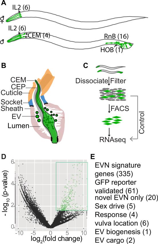
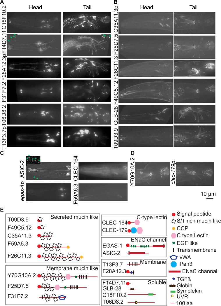
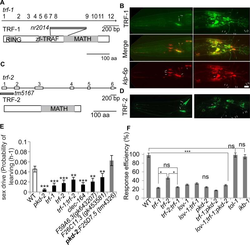
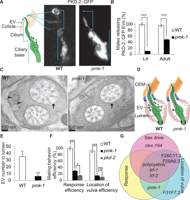
Cilia and extracellular vesicles (EVs) are signaling organelles [1]. Cilia act as cellular sensory antennae, with defects resulting in human ciliopathies. Cilia both release and bind to EVs [1]. EVs are sub-micron-sized particles released by cells and function in both short- and long-range intercellular communication. In C. elegans and mammals, the autosomal dominant polycystic kidney disease (ADPKD) gene products polycystin-1 and polycystin-2 localize to both cilia and EVs, act in the same genetic pathway, and function in a sensory capacity, suggesting ancient conservation [2]. A fundamental understanding of EV biology and the relationship between the polycystins, cilia, and EVs is lacking. To define properties of a ciliated EV-releasing cell, we performed RNA-seq on 27 GFP-labeled EV-releasing neurons (EVNs) isolated from adult C. elegans. We identified 335 significantly overrepresented genes, of which 61 were validated by GFP reporters. The EVN transcriptional profile uncovered new pathways controlling EV biogenesis and polycystin signaling and also identified EV cargo, which included an antimicrobial peptide and ASIC channel. Tumor-necrosis-associated factor (TRAF) homologs trf-1 and trf-2 and the p38 mitogen-activated protein kinase (MAPK) pmk-1 acted in polycystin-signaling pathways controlling male mating behaviors. pmk-1 was also required for EV biogenesis, independent of the innate immunity MAPK signaling cascade. This first high-resolution transcriptome profile of a subtype of ciliated sensory neurons isolated from adult animals reveals the functional components of an EVN.


Figures

Figure 1
Figure 1. Cell-type RNAseq to define the EVN transcriptome
(A) The ciliated EVNs, six in the hermaphrodite (top) and 27 in male (bottom). (B) The male cephalic sensillum. Glial sheath and socket cells form a continuous lumen surrounding the CEM neuronal cilium, which is exposed to the environment directly through a cuticular opening. The lumen is shared by CEM and CEP neurons. EVs are observed in the lumen. (C) Schematic illustration of the differential RNAseq experiment. EVNs were FACS purified from dissociated and filtered cells isolated from synchronized klp-6p::GFP expressing young adult males and hermaphrodites, followed by mRNA extraction, library construction, and RNAseq. Refer to Supplemental Figure 1 for heat map, principal component analysis, and downsampling in silico. (D) Volcano plot showing 335 genes differentially overrepresented in sorted EVNs (colored and boxed in green) compared to whole worms at a false discovery rate of 10%. (E) 335 EVN gene signature genes were validated by GFP expression pattern and functional analysis (for available mutants). The number of genes for each category is listed in parentheses. Refer to Supplemental Tables S1 and S2 for full list of 335 signature genes and original DEseq data, respectively. See Supplemental Table S3 for GO Analysis of EVN overrepresented genes.
Figure 2
Figure 2. EVN signature genes were expressed in all 27 EVNs (A), the 21 polycystin-expressing EVNs (B), or a subset thereof (C, D)
(A) Head IL2 and CEM (left) and tail HOB and RnB (right) images of GFP reporters that were expressed exclusively in all 27 EVNs. (B) A subset of EVN signature genes showed expression in only the male-specific head CEM (left) and tail HOB and RnB (right) neurons. In a and b, arrowheads point to cell bodies in the head and to ray dendrites in the tail. Scale bar, 10 μm. (C) asic-2 was expressed in the six IL2 neurons, egas-1 in four IL2 quadrant (IL2Q) neurons, clec-164 and F59A6.3 in CEM neurons. (D) Y70G10A.2 was expressed only in the HOB EVN and clec-179 in HOB and a few ray EVNs. In all panels, translational GFP reporters are shown by gene name or open reading frame number, transcriptional (promoter) GFP reporters are shown by gene name or ORF followed by p. For translational reporters, arrows point to ciliary localization. In A and C, dashed line boxes were 4X scaled with enhanced contrast and brightness to visualize EVs (green arrows). F14D7.11::GFP localized in cilia and secreted EVs from all 27 EVNs and ASIC-2::GFP localized in IL2 cilia and secreted EVs. (E) Domain analysis of validated genes. Signal peptide sequences were predicted by SignalP 4.1 Server (http://www.cbs.dtu.dk/services/SignalP/). Mucin-domain containing proteins are predicted to be secreted and extensively O-glycosylated in the Serine (S) or Threonine (T) rich domain. Transmembrane domains were predicted by the TM Pred server (http://www.ch.embnet.org/software/TMPRED_form.html). CCP, C-type lectin (CLEC), Pan3, TGFβ, EGF-like, DEG/ENaC channel, vWA (Von Willebrand factor type A), globin, and UVR domains were predicted by Wormbase. Refer to Supplemental Table S4 listing EVN signature genes with predicted or demonstrated function in stress response or innate immunity.
Figure 3
Figure 3. trf-1 and trf-2 coexpressed in male-specific EVNs and acted in the polycystin-signaling pathways
(A, C) Schematic of trf-1 and trf-2 genomic and protein structure, with molecular lesions indicated in genomic structure. (A) The trf-1 locus encodes all common domains shared by mammalian TRAFs: a RING finger domain that may act as an E3 ubiquitin-ligase; a TRAF zinc finger and MATH domain that may mediate protein-protein interaction or oligomerization. The trf-1 (nr2041) mutation deletes part of the zf-TRAF domain and part of the MATH domain and may affect mRNA splicing. (C) TRF-2 has only a MATH domain. The trf-2(tm5167) deletion removes upstream of first exon to the end of the first intron. (B) trf-1 was co-expressed with klp-6 in male-specific, but not IL2, EVNs. (D) TRF-2::GFP was expressed and localized in the same pattern as TRF-1::GFP. (E) TRAF, lectins, and adhesion molecules were required for male sex drive. The tm4326 deletion allele of F25D7.5 suppresses the sex drive defect of pkd-2 mutant males (in bold). The probability of the male leaving the food lawn per hour PL(h-1) is indicated on the Y-axis, the male's genotype on the X-axis. Data was analyzed by Kruskal-Wallis test. * p<0.05, ** p<0.01, *** p<0.001. (F) Response Efficiency (RE) of lov-1, pkd-2, trf-1, trf-2 and Toll pathway genes tol-1 and ikb-1. Data was analyzed with Fisher's exact test, Bonferroni-Holm corrected. * p<0.05, *** p<0.001. ns not significant. The minimum number (n) of individual males analyzed per genotype was 37 for sex drive and 59 for response behavior.
Figure 4
Figure 4. The p38 MAPK pmk-1 was required for EV biogenesis and polycystin-mediated response and vulva location behaviors
(A) PKD-2::GFP CEM ciiary localization in wild-type and pmk-1 males. Cartoon shows the CEM cilium protruding from the cuticular pore and EVs surrounding the cuticle. In wild type, PKD-2::GFP localized to the ciliary base, cilium, and EVs released outsjde the male's nose. Note in wild type, EVs accumulated and surrounded the ciliary pore and cuticle. In pmk-1 males PKD-2::GFP localized normally to the ciliary base and cilium, but was absent from EVs. To show the full length of PKD-2::GFP labeled cilia and the EVs, images of wild type and pmk-1 males were taken with different exposure times, and adjusted with different brightness and contrast. The original images were enlarged six times by the Photoshop CS5 image transformation tool. Scale bar = 1 μm (B) pmk-1 mutant males were defective in PKD-2::GFP labeled EV release in both L4 (n=20 for wild-type, n=29 for pmk-1) and adult stage (n=52 for wild-type and n=43 for pmk-1). Data was analyzed with Fisher's exact test, Bonferroni-Holm corrected; *** p<0.001. (C, D) TEM and cartoon of wild-type and pmk-1 cephalic sensillum at the level of the ciliary transition zone. In wild type, EVs were present in the extracellular lumen. In pmk-1 males, EVs numbers are significantly reduced in the cephalic lumen. In pmk-1 males, the CEM axoneme contained eight rather then nine doublet microtubules of wild-type males. Black arrowheads = EVs, white arrows = CEM transition zone, black arrows = CEP transition zone. Scale bar = 100nm. (E) Bar graph of number of EVs (mean + SEM) in wild-type and pmk-1. Mann-Whitney test was used for statistical analysis; n=7 cephalic sensillae for wild type from two worms, n=4 cephalic sensillae for pmk-1 from one worm. **p=0.01. (F) pmk-1 mutant males are response and Lov defective. Statistical analysis was done by Fisher's exact test, Bonferroni-Holm corrected for response efficiency, RE (n=184 for wild type, 60 for pmk-1 and 264 for pkd-2), and one-way ANOVA for location of vulva efficiency, LE (n=20 for wild type, 20 for pmk-1 and 18 for pkd-2). * p<0.05, *** p<0.001. ns = not significant. (F) The C. elegans polycystins function in a multi-layered signaling network. Venn diagram showing polycystin-mediated mating behaviors are genetically separable. lov-1, pkd-2, and the TRAFs played a central role in sex drive, response, and vulva location while other genes act in specific behavioral pathways. Refer to Supplemental Table S5 for RE and LE of mutants corresponding to new EVN signature genes and pathways.

References

    1. Wood CR, Rosenbaum JL. Ciliary ectosomes: transmissions from the cell's antenna. Trends Cell Biol. 2015;25:276–285. - PMC - PubMed
    1. O'Hagan R, Wang J, Barr MM. Mating behavior, male sensory cilia, and polycystins in Caenorhabditis elegans. Semin Cell Dev Biol. 2014;33:25–33. - PMC - PubMed
    1. Hogan MC, Manganelli L, Woollard JR, Masyuk AI, Masyuk TV, Tammachote R, Huang BQ, Leontovich AA, Beito TG, Madden BJ, Charlesworth MC, Torres VE, LaRusso NF, Harris PC, Ward CJ. Characterization of PKD protein- positive exosome-like vesicles. J Am Soc Nephrol. 2009;20:278–288. - PMC - PubMed
    1. Wood CR, Huang K, Diener DR, Rosenbaum JL. The cilium secretes bioactive ectosomes. Curr Biol. 2013;23:906–911. - PMC - PubMed
    1. Pampliega O, Orhon I, Patel B, Sridhar S, Diaz-Carretero A, Beau I, Codogno P, Satir BH, Satir P, Cuervo AM. Functional interaction between autophagy and ciliogenesis. Nature. 2013;502:194–200. - PMC - PubMed

Publication types