

Demodicosis caused by Demodex canis and Demodex cornei in dogs
- PMID: 26688632
- PMCID: PMC4675583
- DOI: 10.1007/s12639-013-0405-3
Demodicosis caused by Demodex canis and Demodex cornei in dogs
Abstract
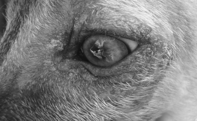
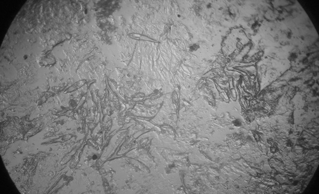
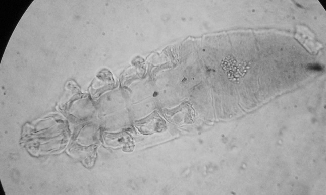
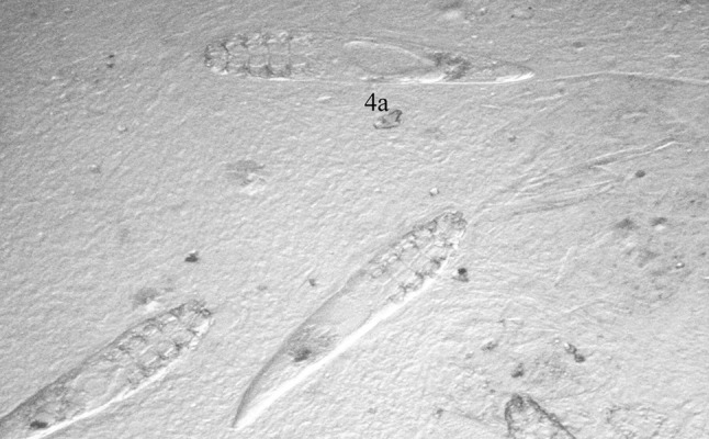
Two mongrel dogs aged between 7 and 9 months in a same house were presented to the clinics with a history of chronic dermatitis associated with pruritus. Clinical examination revealed presence of primary and secondary skin lesions on the face, around the ears, chin, neck, fore limbs and lateral abdomen. Examination of skin scrapings revealed Demodex cornei (majority) and D. canis (minority) in both the dogs. By using hair pluck examination D. canis were detected and by tape impression smears examination large number of adult short-tail Demodex mites were found. D. cornei was identified by based on the morphological characters including short opisthosoma with blind and round terminal end. Mean length of total body, opisthosoma of both types of the mites were differed statistically significant (P < 0.01) but gnathosoma and podosoma did not differ significantly (P > 0.05). Dogs were treated with daily oral ivermectin @ 500 μg/kg/day, external application of amitraz along with supportive therapy. After completion of 45 days of therapy dogs were recovered completely without any side effects.
Keywords: Demodex canis; Demodex cornei; Demodicosis; Dogs.
Figures
References
-
- Hillier A, Desch CE. A new species of Demodex mite in the dog: a case report. Tennessee: Annual Members Meeting of the American Academy of Veterinary Dermatology and the American College of Veterinary Dermatology Nashville; 1997. pp. 118–119.
-
- Lopez R, Reyero D, Banson (2011) First report of canine demodicosis by short-bodied Demodex mite in Spain Rev. Inbero-Latinoam. Parasitology 70(2):219–224
LinkOut - more resources
Full Text Sources
Other Literature Sources