. 2015 Dec 18:11:55.
doi: 10.1186/s13007-015-0098-y. eCollection 2015.

Fast virtual histology using X-ray in-line phase tomography: application to the 3D anatomy of maize developing seeds

Affiliations

Fast virtual histology using X-ray in-line phase tomography: application to the 3D anatomy of maize developing seeds

David Rousseau et al. Plant Methods. .

Abstract

Background: Despite increasing demand, imaging the internal structure of plant organs or tissues without the use of transgenic lines expressing fluorescent proteins remains a challenge. Techniques such as magnetic resonance imaging, optical projection tomography or X-ray absorption tomography have been used with various success, depending on the size and physical properties of the biological material.

Results: X-ray in-line phase tomography was applied for the imaging of internal structures of maize seeds at early stages of development, when the cells are metabolically fully active and water is the main cell content. This 3D imaging technique with histology-like spatial resolution is demonstrated to reveal the anatomy of seed compartments with unequalled contrast by comparison with X-ray absorption tomography. An associated image processing pipeline allowed to quantitatively segment in 3D the four compartments of the seed (embryo, endosperm, nucellus and pericarp) from 7 to 21 days after pollination.

Conclusion: This work constitutes an innovative quantitative use of X-ray in-line phase tomography as a non-destructive fast method to perform virtual histology and extends the developmental stages accessible by this technique which had previously been applied in seed biology to more mature samples.

Keywords: Image segmentation; Virtual histology; X ray in-line phase tomography; maize seeds; plant development.


Figures
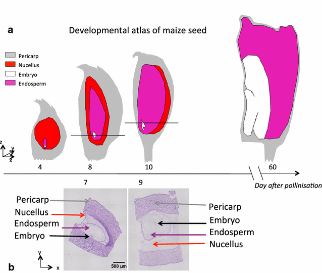
Fig. 1
Fig. 1
Developmental atlas of maize seed. a is a representation of maize seed development under the form of 2D drawings. b shows a conventional destructive histology pictures at 7 and 9 days after pollination (DAP). The level of the slice where these two pictures were extracted is indicated by the black horizontal line in the corresponding drawing in a
Fig. 2
Fig. 2
Maize seeds imaging by conventional absorption X-ray tomography without and with contrast agents. 12 DAP (Days After Pollination) old maize ear imaged by conventional absorption X-ray tomography and treated without contrast agents (a and b) or with Gadolinium (c and d) or Iobitridol (e and f) contrast agents during 24h prior to imaging. a and c represent transversal section of ear, whereas B and D represent longitudinal sections. 3-D reconstruction of the maize ear (e), and its zoom (f) in which only the brightest pixels are displayed, showing high X-ray contrast in ear vasculature and base of kernel
Fig. 3
Fig. 3
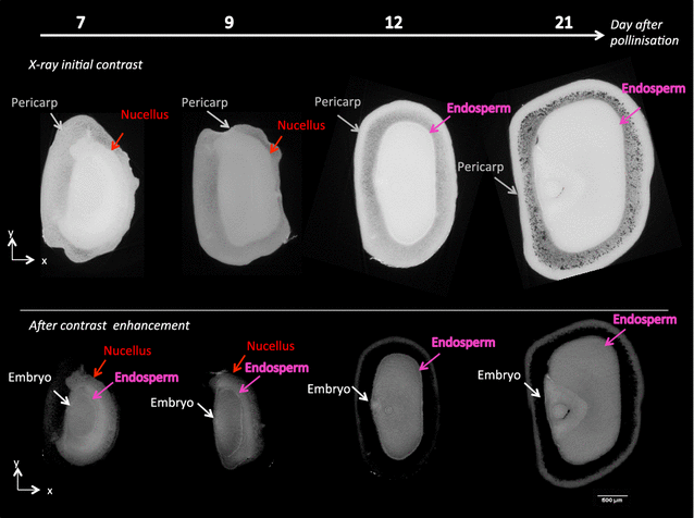
X-ray in-line phase tomography of maize seeds at 4 dates after pollination. Top line initial contrast enhancement on the whole gray level dynamic. Bottom line alternative contrast after dynamic reduction
Fig. 4
Fig. 4
3D segmentation of maize seeds at 4 different developemental stages corresponding to 7, 9, 12 and 21 days after pollination. The segmentation represented under two different angles of view are to be compared with the usual 2D respresentation in Fig. 1. The colors correspond to grey (pericarp), red (nucellus), pink (endosperm), white (embryo). A video of these segmentations is available Additional file 1.
Fig. 5
Fig. 5
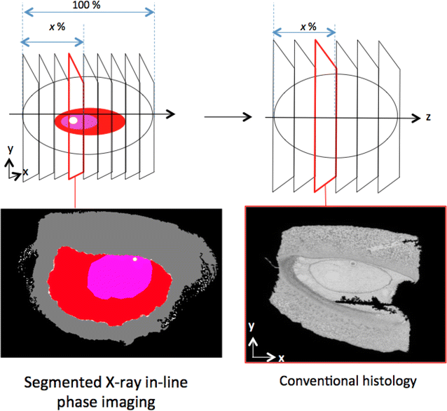
Comparison of a virtual slice obtained from X-ray in-line phase tomography with a real slice obtained from conventional histology. As illustrated in upper panel, slices were taken at the same location in maize seeds both taken at 7 DAP. For X-ray in-line phase image, the colors correspond to pericarp (grey), nucellus (red), endosperm (pink) and embryo (white). The length of the pericarp along the X-axis is taken as the reference (100 %) and the x % correspond to the position of the slice
Fig. 6
Fig. 6
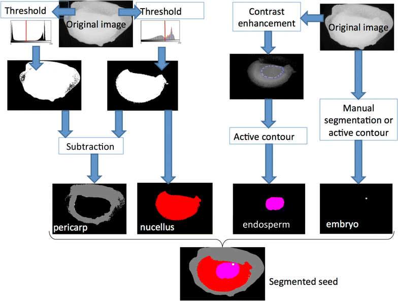
Image processing pipeline developed for the segmentation of the different compartements of maize seed with X-ray in-line phase tomography

References

    1. Fischer RS, Wu Y, Kanchanawong P, Shroff H, Waterman CM. Microscopy in 3D: a biologist’s toolbox. Trends Cell Biol. 2011;21(12):682–691. doi: 10.1016/j.tcb.2011.09.008. - DOI - PMC - PubMed
    1. Dean RJ, Stait-Gardner T, Clarke SJ, Rogiers SY, Bobek G, Price WS. Use of diffusion magnetic resonance imaging to correlate the developmental changes in grape berry tissue structure with water diffusion patterns. Plant methods. 2014;10(1):35. doi: 10.1186/1746-4811-10-35. - DOI - PMC - PubMed
    1. Metzner R, Eggert A, van Dusschoten D, Pflugfelder D, Gerth S, Schurr U, Uhlmann N, Jahnke S. Direct comparison of mri and X-ray CT technologies for 3D imaging of root systems in soil: potential and challenges for root trait quantification. Plant Methods. 2015;11(1):17. doi: 10.1186/s13007-015-0060-z. - DOI - PMC - PubMed
    1. Jahnke S, Menzel MI, Van Dusschoten D, Roeb GW, Bühler J, Minwuyelet S, Blümler P, Temperton VM, Hombach T, Streun M. Combined mri-pet dissects dynamic changes in plant structures and functions. Plant J. 2009;59(4):634–644. doi: 10.1111/j.1365-313X.2009.03888.x. - DOI - PubMed
    1. Suuronen J-P, Peura M, Fagerstedt K, Serimaa R. Visualizing water-filled versus embolized status of xylem conduits by desktop X-ray microtomography. Plant Methods. 2013;9(1):11. doi: 10.1186/1746-4811-9-11. - DOI - PMC - PubMed