Review
. 2015:2015:180972.
doi: 10.1155/2015/180972. Epub 2015 Nov 25.

Optic Disc and Optic Cup Segmentation Methodologies for Glaucoma Image Detection: A Survey

Affiliations
Review

Optic Disc and Optic Cup Segmentation Methodologies for Glaucoma Image Detection: A Survey

Ahmed Almazroa et al. J Ophthalmol. 2015.

Abstract

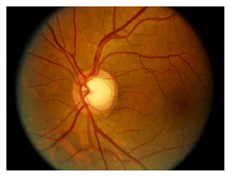
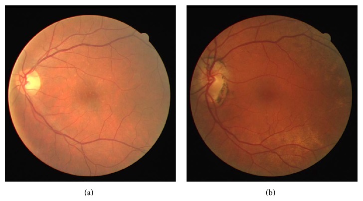
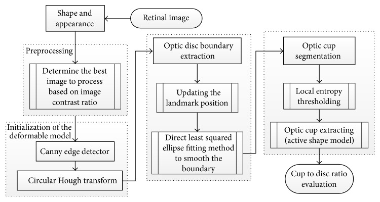
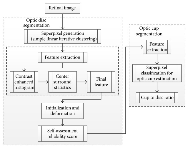
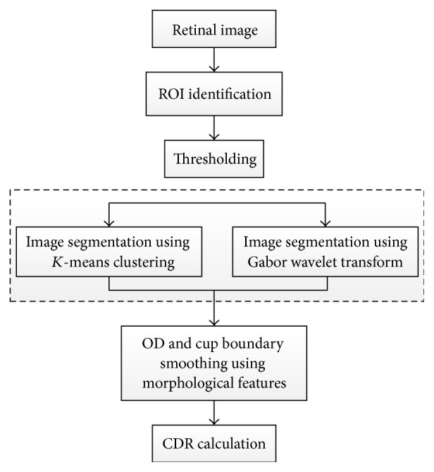
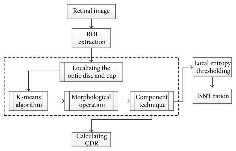
Glaucoma is the second leading cause of loss of vision in the world. Examining the head of optic nerve (cup-to-disc ratio) is very important for diagnosing glaucoma and for patient monitoring after diagnosis. Images of optic disc and optic cup are acquired by fundus camera as well as Optical Coherence Tomography. The optic disc and optic cup segmentation techniques are used to isolate the relevant parts of the retinal image and to calculate the cup-to-disc ratio. The main objective of this paper is to review segmentation methodologies and techniques for the disc and cup boundaries which are utilized to calculate the disc and cup geometrical parameters automatically and accurately to help the professionals in the glaucoma to have a wide view and more details about the optic nerve head structure using retinal fundus images. We provide a brief description of each technique, highlighting its classification and performance metrics. The current and future research directions are summarized and discussed.


Figures

Figure 1
Figure 1
Optic disc in fundus image [14].
Figure 2
Figure 2
Retinal images from DRIVE: (a) normal image, (b) pathological image.
Figure 3
Figure 3
The relation between the ground truth and automatically marked area [9].
Figure 4
Figure 4
Measurement of cup-to-disc ratio for a tilted disc [44].
Figure 5
Figure 5
Measurement of the ISNT rule [45].
Figure 6
Figure 6
Flowchart for algorithm proposed in [39].
Figure 7
Figure 7
Flowchart for algorithm proposed in [31].
Figure 8
Figure 8
Flowchart for algorithm proposed in [32].
Figure 9
Figure 9
ODP determination. ((a), (b), and (c)) Original images. ((a1), (b1), and (c1)) OD pixels provided by the maximum difference method. ((a2), (b2), and (c2)) OD pixels provided by the maximum variance method. ((a3), (b3), and (c3)) OD pixels provided by the low-pass filter method. ((a4), (b4), and (c4)) Final ODP determination.
Figure 10
Figure 10
The calculation process of the circular OD boundary approximation. (R) Red channel. (G) Green channel. ((R1) and (G1)) Vessel elimination. ((R2) and (G2)) Gradient magnitude image. ((R3) and (G3)) Binary image. ((R4) and (G4)) Cleaner version of the binary image. ((R5) and (G5)) Circular OD boundary approximation.
Figure 11
Figure 11
Flowchart for algorithm proposed in [33].
Figure 12
Figure 12
Flowchart for algorithm proposed in [29].
Figure 13
Figure 13
Flowchart for algorithm proposed in [34].
Figure 14
Figure 14
Optic disc segmentation using the proposed method (red), level set method (blue), FCM method (black), CHT method (cyan), and ground truth (green).
Figure 15
Figure 15
Flowchart for algorithm proposed in [35].
Figure 16
Figure 16
(a) The results (blue: without EF, red: with EF, and green: ground truth). (b) The results (cyan: before β-PPA detection, magenta: after β-PPA detection, red: with ellipse correction, and green: ground truth).
Figure 17
Figure 17
Flowchart for algorithm proposed in [30].
Figure 18
Figure 18
Flowchart for algorithm proposed in [37].
Figure 19
Figure 19
Flowchart for algorithm proposed in [38].
Figure 20
Figure 20
Flowchart for algorithm proposed in [36].
Figure 21
Figure 21
Flowchart for algorithm proposed in [17].
Figure 22
Figure 22
Flowchart for algorithms proposed in [28, 40].
Figure 23
Figure 23
Representative results.
Figure 24
Figure 24
Flowchart for algorithm proposed in [55].
Figure 25
Figure 25
Flowchart for algorithm proposed in [66].
Figure 26
Figure 26
Flowchart for algorithm proposed in [59].
Figure 27
Figure 27
Flowchart for algorithm proposed in [63].
Figure 28
Figure 28
OD segmentation using proposed method (red), level set method (blue), and FCM method (black) with ground truth (green).
Figure 29
Figure 29
Optic cup segmentation using the proposed method (blue), ASM method without vessel removal (red), and level set method (black) with ground truth (green).
Figure 30
Figure 30
Flowchart for algorithm proposed in [6].
Figure 31
Figure 31
Sample results of the optic disc. From left to right columns: (a) the original images, (b) the manual “ground truth,” and ((c)–(g)) outlines by the MCV, CHT-ASM, EHT, and MDM.
Figure 32
Figure 32
Sample results of the optic cup. From left to right columns: (a) the original images, (b) the manual “ground truth,” and ((c)–(e)) outlines by the proposed method before ellipse fitting.
Figure 33
Figure 33
Flowchart for algorithm proposed in [62].
Figure 34
Figure 34
Flowchart for algorithm proposed in [58].
Figure 35
Figure 35
Flowchart for algorithm proposed in [65].
Figure 36
Figure 36
Flowchart for algorithm proposed in [61].
Figure 37
Figure 37
Flowchart for algorithm proposed in [60].
Figure 38
Figure 38
Flowchart for algorithm proposed in [42].
Figure 39
Figure 39
Flowchart for algorithm proposed in [64].

References

    1. Quigley H. A., Broman A. T. The number of people with glaucoma worldwide in 2010 and 2020. The British Journal of Ophthalmology. 2006;90(3):262–267. doi: 10.1136/bjo.2005.081224. - DOI - PMC - PubMed
    1. Costagliola C., dell'Omo R., Romano M. R., Rinaldi M., Zeppa L., Parmeggiani F. Pharmacotherapy of intraocular pressure: part I. Parasympathomimetic, sympathomimetic and sympatholytics. Expert Opinion on Pharmacotherapy. 2009;10(16):2663–2677. doi: 10.1517/14656560903300103. - DOI - PubMed
    1. Costagliola C., dell'Omo R., Romano M. R., Rinaldi M., Zeppa L., Parmeggiani F. Pharmacotherapy of intraocular pressure—part II. Carbonic anhydrase inhibitors, prostaglandin analogues and prostamides. Expert Opinion on Pharmacotherapy. 2009;10(17):2859–2870. doi: 10.1517/14656560903300129. - DOI - PubMed
    1. European Glaucoma Society. Terminology and Guidelines for Glaucoma. 4th. Savona, Italy: PubliComm; 2014.
    1. Stewart W. C. Clinical Practice of Glaucoma. Thorofare, NJ, USA: SLACK Incorporated; 1990.