. 2015 Sep 1;1(3):115-123.
doi: 10.1007/s40610-015-0020-4. Epub 2015 Jul 10.

Myoepithelial Cells: Their Origin and Function in Lacrimal Gland Morphogenesis, Homeostasis, and Repair

Affiliations

Myoepithelial Cells: Their Origin and Function in Lacrimal Gland Morphogenesis, Homeostasis, and Repair

Helen P Makarenkova et al. Curr Mol Biol Rep. .

Abstract

Lacrimal gland (LG) is an exocrine tubuloacinar gland that secretes the aqueous layer of the tear film. LG epithelium is composed of ductal, acinar, and myoepithelial cells (MECs) bordering the basal lamina and separating the epithelial layer from the extracellular matrix. Mature MECs have contractile ability and morphologically resemble smooth muscle cells; however, they exhibit features typical for epithelial cells, such as the presence of specific cytokeratin filaments. Increasing evidence supports the assertion that myoepithelial cells (MECs) play key roles in the lacrimal gland development, homeostasis, and stabilizing the normal structure and polarity of LG secretory acini. MECs take part in the formation of extracellular matrix gland and participate in signal exchange between epithelium and stroma. MECs have a high level of plasticity and are able to differentiate into several cell lineages. Here, we provide a review on some of the MEC characteristics and their role in LG morphogenesis, maintenance, and repair.

Keywords: Branching; Epithelium; Fibroblast growth factor; Lacrimal gland; Mesenchyme; Morphogenesis; Myoepithelial cells; Progenitor cells; Regeneration; Smooth muscle actin; Stem.


Conflict of interest statement

Conflict of Interest Helen P. Makarenkova and Darlene A. Dartt declare that they have no conflict of interest.

Figures

Fig. 1
Fig. 1
Lacrimal gland epithelial cell types. The LG epithelium (a) is composed of three major cell types: acinar (b), ductal (c), and myoepithelial (MECs) (d red) cells. Acinar cells synthesize and secrete proteins, water, and electrolytes. Ductal cells modify the secretory fluid by secreting electrolytes and water. Myoepithelial cells (MECs) produce basal membrane proteins and have contractile function
Fig. 2
Fig. 2
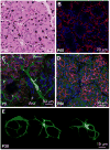
Lacrimal gland myoepithelial cells. (a) Paraffin section of a mouse LG at postnatal day 60 (P60) stained with hematoxylin/eosin (H&E), demonstrating the acinar structure of the gland. MECs (white arrows) are hardly visible. (b) Paraffin section of a mouse LG at P60 stained with the antibody to SMA. Due to the large size of adult MECs, only part of cells can be seen (red) (nuclei are stained with DAPI). (c) Whole mount imaging of the mouse LG at P5. MECs (red) are located around differentiating acini, but not around the main duct (white arrow). Blood vessels (green) are stained with the CD31 antibody; the nuclei are stained with DAPI. (d) Whole mount preparation of a mouse LG at P60 demonstrating large MECs (red) with several long processes around the secretory acini. Epithelial cells are stained with the Panx1 antibody (green), nuclei are stained with DAPI. (e) Typical shapes of MECs in adult LGs as visualized with SMA expression. The cells have 4–7 long processes that may also be branched
Fig. 3
Fig. 3
Lacrimal gland epithelial cell lineage(s). Illustration of the three main hypotheses in cell lineage hierarchy in the LG. (a) Hypothesis I: slow cycling LG stem cell produce a common progenitor that gives rise to all LG lineages, epithelial (acinar and ductal) and myoepithelial (MEC). Hypothesis II: The LG has lineage-restricted stem/progenitor cells that give rise to specific cell types within each lineage. (b) Hypothesis III: This hypothesis suggests that multipotent stem/progenitor cells exist in one of the LG cell lineages. Based on reported plasticity of MECs, MEC lineage may likely contain multipotent stem-like cells that can restore all epithelial lineages upon LG injury. Dashed arrows label unknown connections and straight arrows reported connections

References

    1. Gudjonsson T, Adriance MC, Sternlicht MD, Petersen OW, Bissell MJ. Myoepithelial cells: their origin and function in breast morphogenesis and neoplasia. J Mammary Gland Biol Neoplasia. 2005;10:261–72. - PMC - PubMed
    1. Faraldo MM, Teuliere J, Deugnier MA, Taddei-De La Hosseraye I, Thiery JP, Glukhova MA. Myoepithelial cells in the control of mammary development and tumorigenesis: data from genetically modified mice. J Mammary Gland Biol Neoplasia. 2005;10:211–9. - PubMed
    1. Avci A, Gunhan O, Cakalagaoglu F, Gunal A, Celasun B. The cell with a thousand faces: detection of myoepithelial cells and their contributions in the cytological diagnosis of salivary gland tumors. Diagn Cytopathol. 2012;40:220–7. - PubMed
    1. Abe J, Sugita A, Hamasaki M, et al. Scanning electron microscopic observations of the myoepithelial cells of normal and contracting status in the rat harderian gland. Kurume Med J. 1981;28:103–12. - PubMed
    1. Raubenheimer EJ. The myoepithelial cell: embryology, function, and proliferative aspects. Crit Rev Clin Lab Sci. 1987;25:161–93. - PubMed

LinkOut - more resources