Review
. 2016;89(1061):20150859.
doi: 10.1259/bjr.20150859. Epub 2016 Jan 22.

Multidetector CT in emergency radiology: acute and generalized non-traumatic abdominal pain

Affiliations
Review

Multidetector CT in emergency radiology: acute and generalized non-traumatic abdominal pain

Pasquale Paolantonio et al. Br J Radiol. 2016.

Abstract

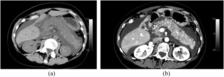
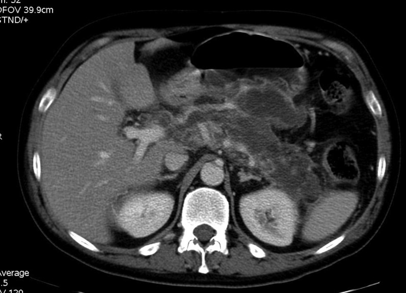
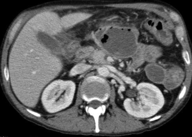
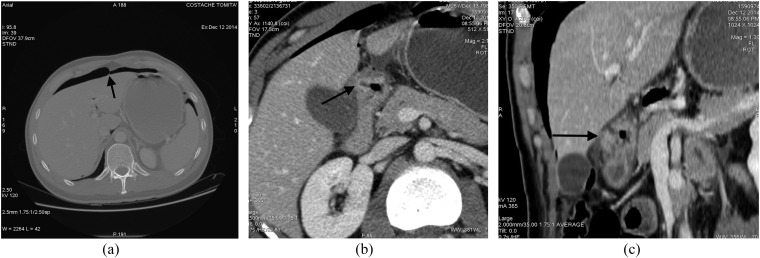
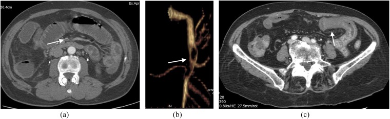
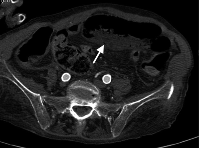
Multidetector CT (MDCT) is an imaging technique that provides otherwise unobtainable information in the diagnostic work-up of patients presenting with acute abdominal pain. A correct working diagnosis depends essentially on understanding the individual patient's clinical data and laboratory findings. In haemodynamically stable patients with acute severe and generalized abdominal pain, MDCT is now the preferred imaging test and gives invaluable diagnostic information, also in unstable patients after stabilization. In this descriptive review, we focus our attention on acute, severe and generalized or undifferentiated non-traumatic abdominal pain. The main differential diagnoses are acute pancreatitis, gastrointestinal perforation, ruptured abdominal aneurysm and acute mesenteric ischaemia. We will provide radiologist readers with a technical guide to optimize MDCT imaging protocols and list the major CT signs essential to reach a correct diagnosis and guide the best treatment.


Figures

Figure 1.
Figure 1.
Multidetector CT study acquired in a patient affected by interstial acute pancreatitis. (a) Unenhanced CT image. (b) Contrast-enhanced CT image. Pancreas is diffusely enlarged and shows low density on unenhanced scan. Pancreatic gland is surrounded by fluid. On contrast-enhanced image, pancreatic gland shows homogeneous enhancement.
Figure 2.
Figure 2.
Multidetector CT study acquired in a patient affected by necrotizing pancreatitis: axial contrast-enhanced CT scan showing diffuse lack of the pancreatic gland enhancement with fluidification of pancreatic tissue as well as peripancreatic fat.
Figure 3.
Figure 3.
A contrast-enhanced CT image acquired in a patient affected by necrotizing pancreatitis and infected walled-off necrosis: CT image shows a capsulated fluid collection in the pancreatic area with internal gas bubbles.
Figure 4.
Figure 4.
Images from a multidetector CT study acquired in a patient affected by perforated duodenal peptic ulcer: (a) axial CT image with wide window setting; (b) contrast-enhanced axial image; (c) an oblique multiplanar reformation. (a) Intraperitoneal free air is clearly visible in the anterior perihepatic space, crossing the midline and accentuating the falciform ligament (arrow). Tiny air bubbles are also visible at the hepatic hilum, close to the caudate lobe and in the sperisplenic space. (b) Thickening of bulbar duodenal wall with deep penetrating ulcer (arrow) is visible. (c) A duodenal wall cleft is better visualized (arrow).
Figure 5.
Figure 5.
Contrast-enhanced multidetector CT images acquired in a patient presenting with ruptured right iliac aneurysm with hemoperitoneum. (a) The site of aneurysm rupture is well depicted (arrow); (b) massive pelvic hemoperitoneum with active extravasation of iodinate contrast medium is shown.
Figure 6.
Figure 6.
Images from multidetector CT (MDCT) study acquired in a patient with rupturing inferior pancreaticoduodenal artery (IPDA) aneurysm. (a) Unenhanced CT image: huge retroperitoneal haematoma is clearly visible in the mesenteric root. (b) Axial contrast-enhanced MDCT image acquired in the early arterial phase; in this image, it is very difficult to identify a tiny aneurysm of a splanchnic arterial vessel (arrow). (c) Three-dimensional volume rendering reconstruction; in this image, a small aneurysm of the IPDA with tiny aneurysm wall bleb is clearly visible (arrow).
Figure 7.
Figure 7.
A coronal multiplanar reformation contrast-enhanced CT images acquired in a patient affected by strangulated small-bowel obstruction and acute mesenteric ischaemia. Ischaemic bowel loops show wall thickening and increased bowel wall enhancement with target appearance.
Figure 8.
Figure 8.
Axial (a) and oblique multiplanar reformation (b) contrast-enhanced CT images acquired in a patient with acute superior mesenteric vein thrombosis.
Figure 9.
Figure 9.
A 55-year-old male patient with acute mesenteric ischaemia due to superior mesenteric artery (SMA) embolization. (a) Arterial embolus as a filling defect (arrow) of SMA on axial contrast-enhanced early arterial phase CT axial image. (b) The extension of SMA embolization is visible on three-dimensional volume rendering selective reconstruction of SMA (arrow). (c) Necrotic changes of bowel wall (arrow) with lack of contrast enhancement.
Figure 10.
Figure 10.
An axial contrast-enhanced CT image acquired in a patient affected by small bowel infarction. Necrotic bowel loop shows dilated lumen, “paper-thin” bowel wall with lack of enhancement. Some vasa recta are gas-filled (arrow).

Similar articles

Cited by

References

    1. Stoker J, van Randen A, Laméris W, Boermeester MA. Imaging patients with acute abdominal pain. Radiology 2009; 253: 31–46. doi: 10.1148/radiol.2531090302 - DOI - PubMed
    1. Bijur P, Latimer C, Gallagher J. Validation of a verbally administered numerical rating scale of acute pain for use in the emergency department. Acad Emerg Med 2003; 10: 390–2. - PubMed
    1. American College of Emergency Physicians (2010). ACEP policy statements: triage scale standardization. Dallas, TX: American College of Emergency Physicians. Cited 1 June 2011. Available from: http://www.acep.org/content.aspx?id=29828&terms=triage%scale
    1. Sala E, Watson CJ, Beadsmoore C, Groot-Wassink T, Fanshawe TR, Smith JC, et al. . A randomized, controlled trial of routine early abdominal computed tomography in patients presenting with non-specific acute abdominal pain. Clin Radiol 2007; 62: 961–9. doi: 10.1016/j.crad.2007.01.030 - DOI - PubMed
    1. Urban BA, Fishman EK. Tailored helical CT evaluation of acute abdomen. Radiographics 2000; 20: 725–49. - PubMed

MeSH terms