. 2015 Dec 21:12:239.
doi: 10.1186/s12974-015-0452-z.

Monomethyl fumarate promotes Nrf2-dependent neuroprotection in retinal ischemia-reperfusion

Affiliations

Monomethyl fumarate promotes Nrf2-dependent neuroprotection in retinal ischemia-reperfusion

Hongkwan Cho et al. J Neuroinflammation. .

Abstract

Background: Retinal ischemia results in neuronal degeneration and contributes to the pathogenesis of multiple blinding diseases. Recently, the fumaric acid ester dimethyl fumarate (DMF) has been FDA-approved for the treatment of multiple sclerosis, based on its neuroprotective and anti-inflammatory effects. Its potential role as a neuroprotective agent for retinal diseases has received little attention. In addition, DMF's mode of action remains elusive, although studies have suggested nuclear factor erythroid 2-related factor 2 (Nrf2) activation as an important mechanism. Here we investigated the neuroprotective role of monomethyl fumarate (MMF), the biologically active metabolite of DMF, in retinal ischemia-reperfusion (I/R) injury, and examined the role of Nrf2 in mediating MMF action.

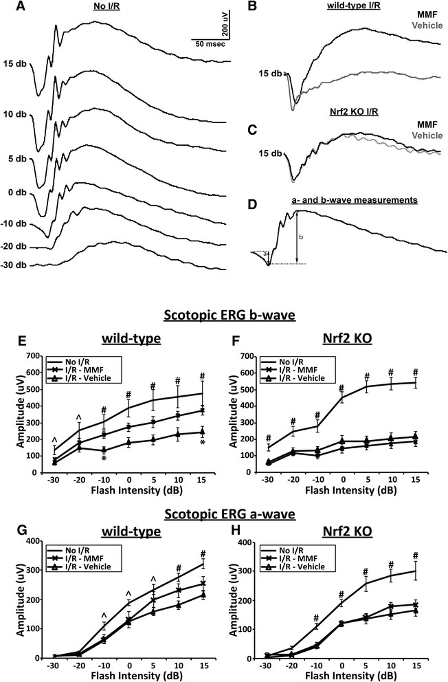
Methods: Wild-type C57BL/6J and Nrf2 knockout (KO) mice were subjected to 90 min of retinal ischemia followed by reperfusion. Mice received daily intraperitoneal injection of MMF. Inflammatory gene expression was measured using quantitative reverse transcription PCR (qRT-PCR) at 48 h after I/R injury. Seven days after I/R, qRT-PCR for Nrf2 target gene expression, immunostaining for Müller cell gliosis and cell loss in the ganglion cell layer (GCL), and electroretinography for retinal function were performed.

Results: The results of this study confirmed that MMF reduces retinal neurodegeneration in an Nrf2-dependent manner. MMF treatment significantly increased the expression of Nrf2-regulated antioxidative genes, suppressed inflammatory gene expression, reduced Müller cell gliosis, decreased neuronal cell loss in the GCL, and improved retinal function measured by electroretinogram (ERG) after retinal I/R injury in wild-type mice. Importantly, these MMF-mediated beneficial effects were not observed in Nrf2 KO mice.

Conclusions: These results indicate that fumaric acid esters (FAEs) exert a neuronal protective function in the retinal I/R model and further validate Nrf2 modulation as a major mode of action of FAEs. This suggests that DMF and FAEs could be a potential therapeutic agent for activation of the Nrf2 pathway in retinal and possibly systemic diseases.


Figures

Fig. 1
Fig. 1
MMF-treated mice exhibit significantly increased Nrf2 target gene expression in I/R retinas in wild-type, but not Nrf2 knockout, mice. qRT-PCR analysis of Nrf2 target genes was performed on retinas harvested 7 days after reperfusion. n = 5-6 mice/group
Fig. 2
Fig. 2
MMF treatment significantly suppresses inflammatory gene expression following I/R in wild-type, but not Nrf2 knockout, mice. qRT-PCR analysis of inflammatory mediators was performed on retinas harvested 48 h after reperfusion. n = 9-10 mice/group
Fig. 3
Fig. 3
MMF treatment significantly reduces retinal Müller cell gliosis associated with I/R injury at day 7. In control eyes, GFAP immunolabeling was mainly present on the ILM (asterisk) (a). In wild-type I/R eyes treated with vehicle, the immunolabeling was significantly (p < 0.001) increased (b) when compared with control eyes and extended almost throughout the entire retina to the ONL in the Müller cell processes (a). With MMF injection, the immunolabeling was significantly (p < 0.05) reduced (b) and mainly extended to the OPL and only a small amount of GFAP immunolabeling was observed in the ONL (a). In Nrf2 KO mice, both vehicle- and MMF-treatment showed extensive GFAP immunofluorescence throughout all retina layers to the ONL (a). I/R-induced Muller cell gliosis was significantly (p < 0.01) higher in Nrf2 KO mice when compared with wild-type mice (b). n = 4-7 mice/group. ILM inner limiting membrane, GCL ganglion cell layer, IPL inner plexiform layer, INL inner nuclear layer, OPL outer plexiform layer, ONL outer nuclear layer
Fig. 4
Fig. 4
MMF treatment protects retinal ganglion cells from I/R-induced cell death in an Nrf2-dependent manner. Representative anti-NeuN stained retinal flat-mount images from WT (a) and KO (c) mice are shown. MMF significantly (p < 0.001) inhibits neuronal cell loss in GCL at 7 days after I/R in WT (b), but not KO (d), mice. n = 4-6 mice/group
Fig. 5
Fig. 5
MMF treatment improves I/R-induced functional impairment in the retina in an Nrf2-dependent manner. An intensity series of dark-adapted flash ERG traces from a representative non-I/R eye (a), and representative traces from I/R eyes with or without MMF treatment to a stimulus flash of 15 dB in wild-type (b) and KO (c) mice are shown. An illustration depicting measurements of a and b wave amplitudes are shown (d). Retinal I/R injury significantly reduces b wave (e and f) and a wave (g and h) amplitudes in both wild-type and KO mice at day 7. With MMF treatment, b wave amplitudes in I/R eyes show significant increases at −10 and 15 dB (e), whereas MMF shows no treatment effect in Nrf2 knockout mice (f). A non-significant trend toward improved a wave amplitude at higher flash intensities of 5, 10, and 15 dB in wild-type mice after MMF treatment (g), whereas no such trend was present in Nrf2 knockout mice (h). *p < 0.05, vehicle I/R compared with MMF I/R; ^p < 0.05, # p < 0.005, No I/R control compared with vehicle I/R. n = 7-10 mice/group

References

    1. Osborne NN, Casson RJ, Wood JP, Chidlow G, Graham M, Melena J. Retinal ischemia: mechanisms of damage and potential therapeutic strategies. Progress in retinal and eye research. 2004;23(1):91–147. doi: 10.1016/j.preteyeres.2003.12.001. - DOI - PubMed
    1. Wei YH, Gong JS, Yoshida T, Eberhart CG, Xu ZH, Kombairaju P, et al. Nrf2 has a protective role against neuronal and capillary degeneration in retinal ischemia-reperfusion injury. Free Radical Bio Med. 2011;51(1):216–24. doi: 10.1016/j.freeradbiomed.2011.04.026. - DOI - PMC - PubMed
    1. Pellegrini-Giampietro DE, Cherici G, Alesiani M, Carla V, Moroni F. Excitatory amino acid release and free radical formation may cooperate in the genesis of ischemia-induced neuronal damage. The Journal of neuroscience : the official journal of the Society for Neuroscience. 1990;10(3):1035–41. - PMC - PubMed
    1. McCord JM. Oxygen-derived free radicals in postischemic tissue injury. The New England journal of medicine. 1985;312(3):159–63. doi: 10.1056/NEJM198501173120305. - DOI - PubMed
    1. Korthuis RJ, Granger DN. Reactive oxygen metabolites, neutrophils, and the pathogenesis of ischemic-tissue/reperfusion. Clinical cardiology. 1993;16(4 Suppl 1):I19–26. doi: 10.1002/clc.4960161307. - DOI - PubMed

Publication types

MeSH terms