

Transport across Schlemm's canal endothelium and the blood-aqueous barrier
- PMID: 26689753
- PMCID: PMC4893895
- DOI: 10.1016/j.exer.2015.11.026
Transport across Schlemm's canal endothelium and the blood-aqueous barrier
Abstract
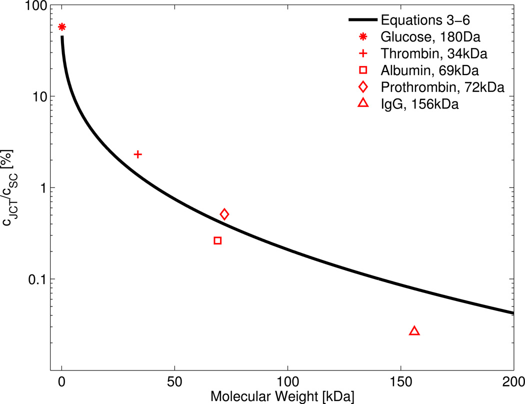
The majority of trabecular outflow likely crosses Schlemm's canal (SC) endothelium through micron-sized pores, and SC endothelium provides the only continuous cell layer between the anterior chamber and episcleral venous blood. SC endothelium must therefore be sufficiently porous to facilitate outflow, while also being sufficiently restrictive to preserve the blood-aqueous barrier and prevent blood and serum proteins from entering the eye. To understand how SC endothelium satisfies these apparently incompatible functions, we examined how the diameter and density of SC pores affects retrograde diffusion of serum proteins across SC endothelium, i.e. from SC lumen into the juxtacanalicular tissue (JCT). Opposing retrograde diffusion is anterograde bulk flow velocity of aqueous humor passing through pores, estimated to be approximately 5 mm/s. As a result of this relatively large through-pore velocity, a mass transport model predicts that upstream (JCT) concentrations of larger solutes such as albumin are less than 1% of the concentration in SC lumen. However, smaller solutes such as glucose are predicted to have nearly the same concentration in the JCT and SC. In the hypothetical case that, rather than micron-sized pores, SC formed 65 nm fenestrae, as commonly observed in other filtration-active endothelia, the predicted concentration of albumin in the JCT would increase to approximately 50% of that in SC. These results suggest that the size and density of SC pores may have developed to allow SC endothelium to maintain the blood-aqueous barrier while simultaneously facilitating aqueous humor outflow.
Keywords: Biological transport phenomena; Blood-aqueous barrier; Diffusion; Glaucoma; Schlemm's canal endothelium.
Copyright © 2016 Elsevier Ltd. All rights reserved.
Figures

References
-
- Aukland K, Reed RK. Interstitial-lymphatic mechanisms in the control of extracellular fluid volume. Physiological reviews. 1993;73:1–78. - PubMed
-
- Barsotti MF, Bartels SP, Freddo TF, Kamm RD. The source of protein in the aqueous humor of the normal monkey eye. Investigative Ophthalmology and Visual Science. 1992;33(3):581–595. - PubMed
-
- Baumeister M, Terzi E, Ekici Y, Kohnen T. Comparison of manual and automated methods to determine horizontal corneal diameter. Journal of Cataract & Refractive Surgery. 2004;30:374–380. - PubMed
-
- Bill A, Svedbergh B. Scanning electron microscopic studies of the trabecular meshwork and the canal of Schlemm. Acta Ophthalmologica. 1972;50:295–320. - PubMed
Publication types
MeSH terms
Grants and funding
LinkOut - more resources
Full Text Sources
Other Literature Sources
Medical
Research Materials
