Review
. 2015 Dec 22:12:193.
doi: 10.1186/s12985-015-0421-2.

Porcine epidemic diarrhea virus: An emerging and re-emerging epizootic swine virus

Affiliations
Review

Porcine epidemic diarrhea virus: An emerging and re-emerging epizootic swine virus

Changhee Lee. Virol J. .

Erratum in

Abstract

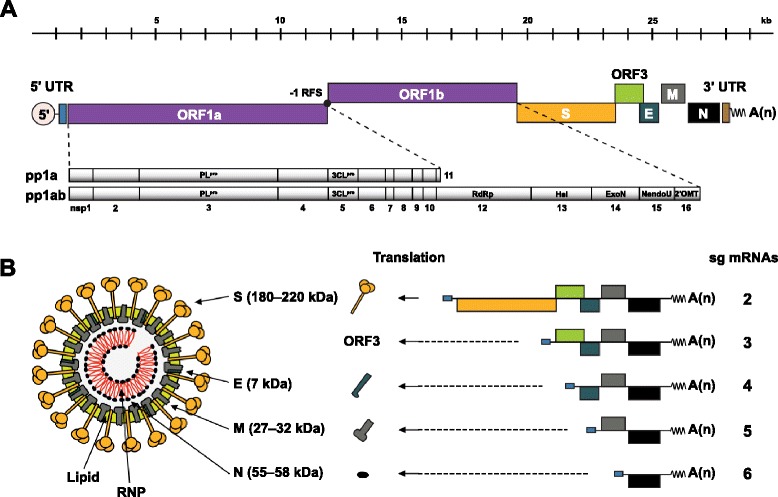
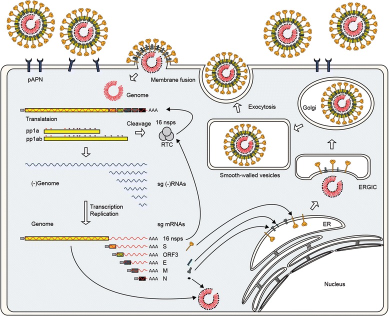
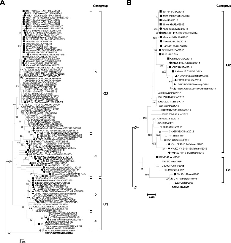
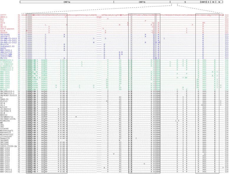
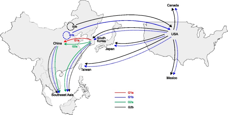
The enteric disease of swine recognized in the early 1970s in Europe was initially described as "epidemic viral diarrhea" and is now termed "porcine epidemic diarrhea (PED)". The coronavirus referred to as PED virus (PEDV) was determined to be the etiologic agent of this disease in the late 1970s. Since then the disease has been reported in Europe and Asia, but the most severe outbreaks have occurred predominantly in Asian swine-producing countries. Most recently, PED first emerged in early 2013 in the United States that caused high morbidity and mortality associated with PED, remarkably affecting US pig production, and spread further to Canada and Mexico. Soon thereafter, large-scale PED epidemics recurred through the pork industry in South Korea, Japan, and Taiwan. These recent outbreaks and global re-emergence of PED require urgent attention and deeper understanding of PEDV biology and pathogenic mechanisms. This paper highlights the current knowledge of molecular epidemiology, diagnosis, and pathogenesis of PEDV, as well as prevention and control measures against PEDV infection. More information about the virus and the disease is still necessary for the development of effective vaccines and control strategies. It is hoped that this review will stimulate further basic and applied studies and encourage collaboration among producers, researchers, and swine veterinarians to provide answers that improve our understanding of PEDV and PED in an effort to eliminate this economically significant viral disease, which emerged or re-emerged worldwide.


Figures

Fig. 1
Fig. 1
Schematic representations of PEDV genome organization and virion structure. a The structure of PEDV genomic RNA. The 5’-capped and 3’-polyadenylated genome of approximately 28 kb is shown at the top. The viral genome is flanked by UTRs and is polycistronic, harboring replicase ORFs 1a and 1b followed by the genes encoding the envelope proteins, the N protein, and the accessory ORF3 protein. S, spike; E, envelope; M, membrane; N, nucleocapsid. Expression of the ORF1a and 1b yields two known polyproteins (pp1a and pp1ab) by −1 programmed RFS, which are co-translationally or post-translationally processed into at least 16 distinct nsps designated nsp1–16 (bottom). PLpro, papain-like cysteine protease; 3CLpro, the main 3C-like cysteine protease; RdRp; RNA-dependent RNA polymerase; Hel, helicase; ExoN, 3’ → 5’ exonuclease; NendoU, nidovirus uridylate-specific endoribonuclease; 2’OMT, ribose-2’-O-methyltransferase. b Model of PEDV structure. The structure of the PEDV virion is illustrated on the left. Inside the virion is the RNA genome associated with the N protein to form a long, helical ribonucleoprotein (RNP) complex. The virus core is enclosed by a lipoprotein envelope, which contains S, E, and M proteins. The predicted molecular sizes of each structural protein are indicated in parentheses. A set of corresponding sg mRNAs (sg mRNA; 2–6), through which canonical structural proteins or nonstructural ORF3 protein are exclusively expressed via a co-terminal discontinuous transcription strategy, are also depicted on the right
Fig. 2
Fig. 2
Overview of the PEDV replication cycle. PEDV binds pAPN via the spike protein. Penetration and uncoating occur after the S protein-mediated fusion of the viral envelope with the plasma membrane. Following disassembly, the viral genome is released into the cytoplasm and immediately translated to yield replicases ppla and pp1ab. These polyproteins are proteolytically cleaved into 16 nsps comprising the replication and transcription complex (RTC) that first engages in the minus-strand RNA synthesis using genomic RNA. Both full- and sg-length minus strands are produced and used to synthesize full-length genomic RNA and sg mRNAs. Each sg mRNA is translated to yield only the protein encoded by the 5’-most ORF of the sg mRNA. The envelope S, E, and M proteins are inserted in the ER and anchored in the Golgi apparatus. The N protein interacts with newly synthesized genomic RNA to form helical RNP complexes. The progeny virus is assembled by budding of the preformed RNP at the ER-Golgi intermediate compartment (ERGIC) and then released by the exocytosis-like fusion of smooth-walled, virion-containing vesicles with the plasma membrane [22]
Fig. 3
Fig. 3
Phylogenetic analyses of global PEDV strains based on nucleotide sequences of the spike genes (a) and full-length genomes (b). A putative similar region of the spike protein and the complete genome sequence of TGEV was included as an outgroup in each panel. Multiple sequence alignments were performed using ClustalX 2.0 program and the phylogenetic tree was constructed from aligned nucleotide sequences using the distance-based neighbor-joining method of MEGA5.2 software. Numbers at each branch represent bootstrap values greater than 50 % of 1000 replicates. Names of the strains, countries and years of isolation, GenBank accession numbers, genogroups, and subgroups are shown. PEDV isolates identified in different countries are indicated by corresponding symbols: Europe (solid triangles), South Korea (sold circles), Thailand and Vietnam, (sold diamonds), and the United States (solid squares). Scale bars indicate nucleotide substitutions per site
Fig. 4
Fig. 4
Amino acid sequence alignment of the N-terminal region of the S protein of global PEDV strains. The top illustration represents the organization of the PEDV genome. Only the corresponding alignment of amino acid sequences of the N-terminal region containing hypervariable regions [26] is shown. Dashes (−) indicate deleted sequences. Potential N glycosylation sites predicted by GlycoMod Tool (http://www.expasy.ch/tools/glycomod/) are shown in boldface type. Genetic subgroups of PEDV were marked with different colors: G1a (red), G1b (blue), G2a (green), and G2b (black). Insertions and deletions (indels) within PEDV isolates compared to the prototype CV777 strain are shaded. Amino acids representing potential hypervariable domains are indicated by solid boxes
Fig. 5
Fig. 5
Potential international PEDV transmission routes. Genetic subgroups of PEDV were marked with different colors as described in the legend to Fig. 4. Solid lines indicate PEDV spreads that have already occurred between countries; dotted lines indicate PEDV spreads that are expected to happen eventually; dashed circular arrows denote genetic mutations or recombination events that lead to the emergence of the novel subtypes

References

    1. Oldham J. Letter to the editor. Pig Farming. 1972;10:72–3.
    1. Chasey D, Cartwright SF. Virus-like particles associated with porcine epidemic diarrhea. Res Vet Sci. 1978;25:255–6. - PMC - PubMed
    1. Wood EN. An apparently new syndrome of porcine epidemic diarrhoea. Vet Rec. 1977;100:243–4. doi: 10.1136/vr.100.12.243. - DOI - PubMed
    1. Pensaert MB, de Bouck P. A new coronavirus-like particle associated with diarrhea in swine. Arch Virol. 1978;58:243–7. doi: 10.1007/BF01317606. - DOI - PMC - PubMed
    1. Debouck P, Pensaert M. Experimental infection of pigs with a new porcine enteric coronavirus, CV777. Am J Vet Res. 1980;41:219–23. - PubMed

Publication types

MeSH terms