

Immunochemical detection of the occupational allergen, methylene diphenyl diisocyanate (MDI), in situ
- PMID: 26690039
- PMCID: PMC4753098
- DOI: 10.1016/j.jim.2015.12.008
Immunochemical detection of the occupational allergen, methylene diphenyl diisocyanate (MDI), in situ
Abstract
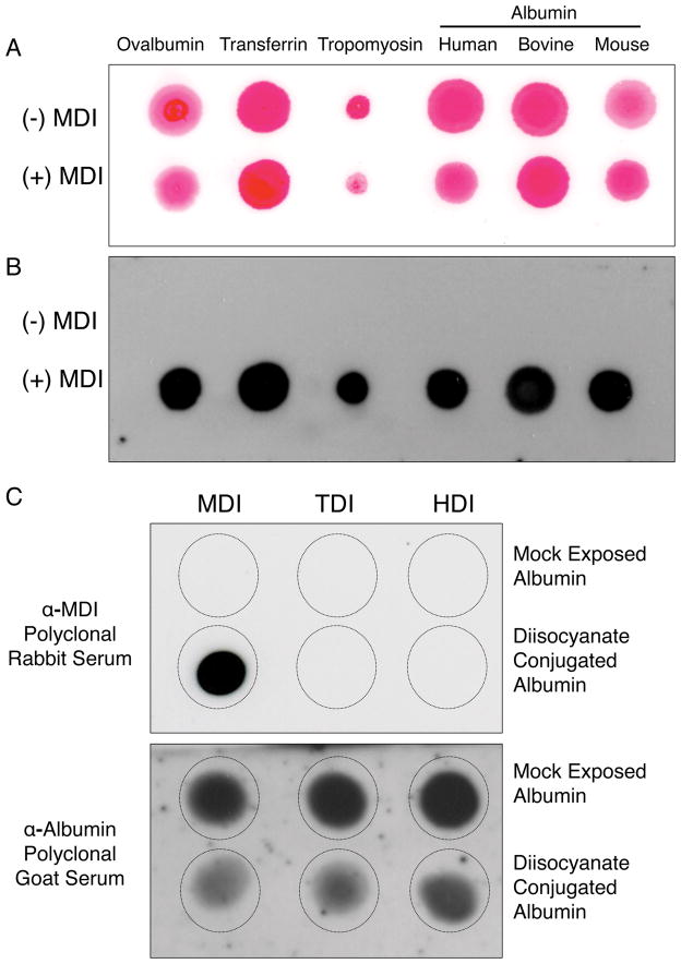
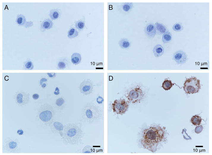
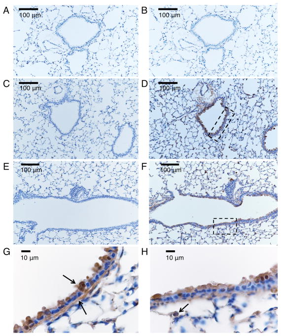
Diisocyanate chemicals essential to polyurethane production are a well-recognized cause of occupational asthma. The pathogenesis of diisocyanate-induced asthma, including the pathways by which the chemical is taken up and its distribution in exposed tissue, especially the lung, remains unclear. We developed an antiserum with specificity for methylene diphenyl diisocyanate (MDI) the most abundantly produced and utilized diisocyanate world-wide, and established its ability to detect MDI in situ. Polyclonal MDI-specific IgG was induced by immunizing rabbits with MDI-conjugated to keyhole limpet hemocyanin (KLH) emulsified in complete Freund's adjuvant, followed by two booster injections with incomplete Freund's adjuvant. The antiserum contains IgG that recognize a variety of different MDI conjugated proteins, but not unconjugated or mock exposed proteins by dot blot analysis. The antiserum further demonstrates specificity for proteins conjugated with MDI, but not other commonly used diisocyanates. Immunochemical studies with cytospun airway cells and formalin-fixed paraffin embedded lung tissue sections from mice intranasally exposed to MDI (as reversibly reactive glutathione conjugates, e.g. GSH-MDI) demonstrated the antiserum's ability to detect MDI in tissue samples. The data demonstrate penetration of MDI into the lower airways, localized deposition in the epithelial region surrounding airways, and uptake by alveolar macrophages. The new immunochemical reagent should be useful for further studies delineating the uptake and tissue distribution of MDI, especially as it relates to adverse health effects from exposure.
Keywords: Airways; Antiserum; Methylene-diphenyl-diisocyanate (MDI); Occupational asthma.
Copyright © 2015 Elsevier B.V. All rights reserved.
Figures
Similar articles
-
Severe asthma and death in a worker using methylene diphenyl diisocyanate MDI asthma death.Am J Ind Med. 2022 Mar;65(3):166-172. doi: 10.1002/ajim.23323. Epub 2022 Jan 14. Am J Ind Med. 2022. PMID: 35028957 Free PMC article.
-
Connecting glutathione with immune responses to occupational methylene diphenyl diisocyanate exposure.Chem Biol Interact. 2013 Sep 5;205(1):38-45. doi: 10.1016/j.cbi.2013.06.005. Epub 2013 Jun 20. Chem Biol Interact. 2013. PMID: 23791970 Free PMC article.
-
Molecular determinants of humoral immune specificity for the occupational allergen, methylene diphenyl diisocyanate.Mol Immunol. 2013 Jun;54(2):233-7. doi: 10.1016/j.molimm.2012.11.017. Epub 2013 Jan 4. Mol Immunol. 2013. PMID: 23295252 Free PMC article.
-
Methylene diphenyl diisocyanate occupational exposure data in industry (1998-2020): A descriptive summary from an industrial hygiene perspective.Toxicol Ind Health. 2023 Aug;39(8):407-420. doi: 10.1177/07482337231176604. Epub 2023 Jun 2. Toxicol Ind Health. 2023. PMID: 37269111 Free PMC article. Review.
-
New mechanisms in diisocyanate-mediated allergy/toxicity: are microRNAs in play?Curr Opin Allergy Clin Immunol. 2025 Apr 1;25(2):75-82. doi: 10.1097/ACI.0000000000001043. Epub 2024 Oct 23. Curr Opin Allergy Clin Immunol. 2025. PMID: 39450940 Free PMC article. Review.
Cited by
-
Severe asthma and death in a worker using methylene diphenyl diisocyanate MDI asthma death.Am J Ind Med. 2022 Mar;65(3):166-172. doi: 10.1002/ajim.23323. Epub 2022 Jan 14. Am J Ind Med. 2022. PMID: 35028957 Free PMC article.
-
Analysis of Lung Gene Expression Reveals a Role for Cl- Channels in Diisocyanate-induced Airway Eosinophilia in a Mouse Model of Asthma Pathology.Am J Respir Cell Mol Biol. 2020 Jul;63(1):25-35. doi: 10.1165/rcmb.2019-0400OC. Am J Respir Cell Mol Biol. 2020. PMID: 32101465 Free PMC article.
-
Molecular Characterization and Experimental Utility of Monoclonal Antibodies with Specificity for Aliphatic Di- and Polyisocyanates.Monoclon Antib Immunodiagn Immunother. 2020 Jun;39(3):66-73. doi: 10.1089/mab.2020.0006. Epub 2020 Apr 17. Monoclon Antib Immunodiagn Immunother. 2020. PMID: 32302507 Free PMC article.
-
Glutathione reactivity with aliphatic polyisocyanates.PLoS One. 2022 Jul 15;17(7):e0271471. doi: 10.1371/journal.pone.0271471. eCollection 2022. PLoS One. 2022. PMID: 35839242 Free PMC article.
-
MicroRNA-mediated calcineurin signaling activation induces CCL2, CCL3, CCL5, IL8, and chemotactic activities in 4,4'-methylene diphenyl diisocyanate exposed macrophages.Xenobiotica. 2021 Dec;51(12):1436-1452. doi: 10.1080/00498254.2021.2005851. Epub 2021 Dec 2. Xenobiotica. 2021. PMID: 34775880 Free PMC article.
References
-
- Allport DC, Gilbert DS, Outterside SM, editors. MDI and TDI: Safety, Health and the Environment: A Source Book and Practical Guide. Chichester Wiley, Wiley; 2003. - DOI
Publication types
MeSH terms
Substances
Grants and funding
LinkOut - more resources
Full Text Sources
Other Literature Sources
