. 2015 Dec 21;10(12):e0145658.
doi: 10.1371/journal.pone.0145658. eCollection 2015.

Longitudinal Cerebral Perfusion Change in Transient Global Amnesia Related to Left Posterior Medial Network Disruption

Affiliations

Longitudinal Cerebral Perfusion Change in Transient Global Amnesia Related to Left Posterior Medial Network Disruption

Jae-Won Jang et al. PLoS One. .

Abstract

Background: The pathophysiology of transient global amnesia (TGA) is not fully understood. Previous studies using single photon emission computed tomography (SPECT) have reported inconclusive results regarding cerebral perfusion. This study was conducted to identify the patterns of regional cerebral blood flow (rCBF) in TGA patients via longitudinal SPECT analysis. An association between the observed SPECT patterns and a pathophysiological mechanism was considered.

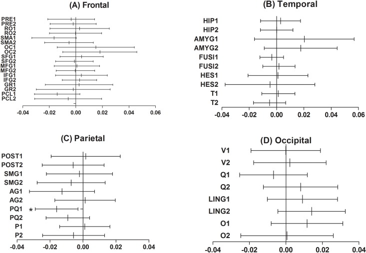
Methods: Based on the TGA registry database of Seoul National University Bundang Hospital, 22 TGA patients were retrospectively identified. The subjects underwent initial Tc-99m-ethyl cysteinate dimer (ECD) SPECT within 4 days of an amnestic event and underwent follow-up scans approximately 6 months later. The difference in ECD uptake between the two scans was measured via voxel-based whole brain analysis, and the quantified ECD uptake was tested using a paired t-test.

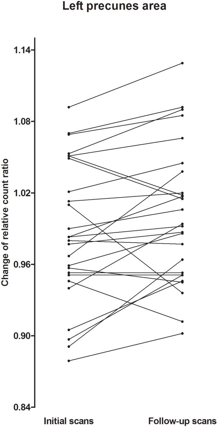
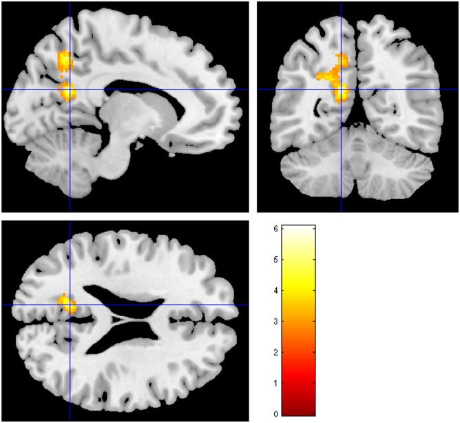
Results: The TGA patients had significantly decreased cerebral perfusion at the left precuneus (P<0.001, uncorrected) and at the left superior parietal and inferior temporal gyrus according to the voxel-based whole brain analysis (P<0.005, uncorrected). A difference in the quantified ECD uptake between the 2 scans was also found at the left precuneus among the 62 cortical volumes of interest (P = 0.018, Cohen's d = -0.25).

Conclusion: We identified left hemispheric lateralized hypoperfusion that may be related to posterior medial network disruption. These findings may be a contributing factor to the pathophysiology of TGA.


Conflict of interest statement

Competing Interests: The authors have declared that no competing interests exist.

Figures

Fig 1
Fig 1. The mean difference in the regional cerebral perfusion between the initial and follow-up stages of TGA.
Only the left precuneus exhibited a significant difference between the 2 scans among the 62 volumes of interest (VOIs) (P = 0.018, Cohen’s d = -0.25). The VOI of the central structures, the insula and the cingulate gyrus, which showed no significant differences between the 2 scans, are not presented here. The mean difference was obtained by subtracting the ECD uptake of the follow-up scan from the initial scans and demonstrated by the VOIs according to the cortical lobes. The vertical line in the center of the each horizontal line represents the mean difference, and the whiskers indicate the 95% confidence interval. Abbreviation: 1,left; 2,right; PRE, precentral gyrus; RO, roalandic operculum; SMA, supplementary motor area; OC, olfactory cortex; SFG, superior frontal gyrus; MFG, middle frontal gyrus; GR, gyrus rectus; PCL, paracentral lobule; HIP, hippocampus; AMYG, amygdala; FUSI, fusiform gyrus; HES, Heschl gyrus; T, temporal gyrus; POST, postcentral gyrus; SMG, supramarginal gyrus; AG, angular gyrus; PQ, precuneus; P, superior and inferior parietal lobule; V, calcarine fissure and surrounding cortex; Q, cuneus; LING, lingual gyrus; O, lateral remainder of occipital lobe.
Fig 2
Fig 2. Changes in ECD uptake counts across the 6-month-interval of the TGA patients at the left precuneus.
The ECD count of each patient was plotted to demonstrate the change between the two scans using a paired Student’s t test.
Fig 3
Fig 3. Statistical parametric mapping projections of areas with significantly reduced cerebral perfusion when comparing the follow-up SPECT scans with the initial scans (Uncorrected p<0.005).
The warm color represents the relative hypoperfusion of the initial scans compared to the follow-up scans.

References

    1. Fisher CM, Adams RD. TRANSIENT GLOBAL AMNESIA. Acta neurologica Scandinavica Supplementum. 1964;40:SUPPL 9:1–83. Epub 1964/01/01. . - PubMed
    1. Sander K, Sander D. New insights into transient global amnesia: recent imaging and clinical findings. 2005;4(7):437–44. 10.1016/S1474-4422(05)70121-6 . - DOI - PubMed
    1. Bartsch T, Alfke K, Deuschl G, Jansen O. Evolution of hippocampal CA-1 diffusion lesions in transient global amnesia. 2007;62(5):475–80. 10.1002/ana.21189 . - DOI - PubMed
    1. Lewis SL. Aetiology of transient global amnesia. Lancet. 1998;352(9125):397–9. Epub 1998/08/26. 10.1016/s0140-6736(98)01442-1 . - DOI - PubMed
    1. Yang Y, Kim S, Kim JH. Ischemic evidence of transient global amnesia: location of the lesion in the hippocampus. Journal of clinical neurology (Seoul, Korea). 2008;4(2):59–66. Epub 2009/06/11. 10.3988/jcn.2008.4.2.59 - DOI - PMC - PubMed

LinkOut - more resources