. 2016 Apr 1:98:124-133.
doi: 10.1016/j.ymeth.2015.12.007. Epub 2015 Dec 12.

Quantitative spatial analysis of transcripts in multinucleate cells using single-molecule FISH

Affiliations

Quantitative spatial analysis of transcripts in multinucleate cells using single-molecule FISH

ChangHwan Lee et al. Methods. .

Abstract

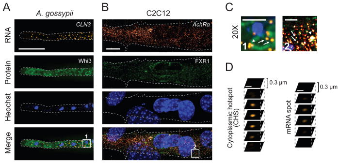
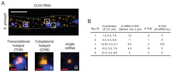
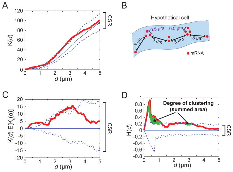
mRNA positioning in the cell is important for diverse cellular functions and proper development of multicellular organisms. Single-molecule RNA FISH (smFISH) enables quantitative investigation of mRNA localization and abundance at the level of individual molecules in the context of cellular features. Details about spatial mRNA patterning at various times, in different genetic backgrounds, at different developmental stages, and under varied environmental conditions provide invaluable insights into the mechanisms and functions of spatial regulation. Here, we describe detailed methods for performing smFISH along with immunofluorescence for two large, multinucleate cell types: the fungus Ashbya gossypii and cultured mouse myotubes. We also put forward a semi-automated image processing tool that systematically detects mRNAs from smFISH data and statistically analyzes the spatial pattern of mRNAs using a customized MATLAB code. These protocols and image analysis tools can be adapted to a wide variety of transcripts and cell types for systematically and quantitatively analyzing mRNA distribution in three-dimensional space.

Keywords: A. gossypii; C2C12 myotubes; Ripley’s H; Single molecule RNA FISH; Spatial analysis.


Figures

Figure 1
Figure 1
smFISH with immunofluorescence (IF) (A) CLN3 mRNA (orange; smFISH) and Whi3-GFP (green; IF) are visualized by smFISH and IF, respectively in A. gossypii. Rabbit anti-GFP IgG (1:100) and rat antirabbit IgG conjugated with Alexa-488 (1:500; Santa Cruz Biotech, TX) are used for IF. (B) AchRα transcripts (orange; smFISH) and FXR1 (green; IF) in differentiated C2C12 mouse myotubes. FXR1 was detected using 1:5,000 rabbit α-FXR1 ML14 antiserum (a generous gift from Edward Khandjian at Universite Laval), and 1:200 goat α-rabbit Alexa 488 (Molecular probes). (A,B) Nuclei are visualized by Hoechst staining. The gray dashed line indicates cell outline. Scale bar: 10 μm. (C) Magnification (20X) of a region within boxes in Figure 1A and 1B, bottom panels. Arrowheads indicate the cytoplasmic hotspots and arrows indicate single mRNA foci. Scale bar: 2 μm. (D) Z-sections of a cytoplasmic hotspot and mRNA spot. Scale bar: 500 nm.
Figure 2
Figure 2
Analysis of smFISH data (A) A transcriptional hotspot (THS), cytoplasmic hotspot (CHS), and single mRNA spot are shown as a maximum-intensity Z-projection. Each white square box is enlarged in the bottom panel. The gray dashed line indicates the cell outline. White scale bar: 5 μm. Red scale bars: 500 nm. (B) An example of output data (Material 5) generated by the customized MATLAB code (Materials 4,6). The coordinates in the table refer to the positions of nuclei in the smFISH image shown in A. The top-left corner of the image represents 0 μm for X and Y coordinates. In the Z-stack of the smFISH image, the bottom Z-slice represents 0 μm for Z coordinate. The MATLAB data includes the following: the number of mRNAs within 2 μm of the center point of each nucleus (i.e. within a spherical ROI with a 2 μm radius), the number of THSs detected within each nucleus, and the number of CHSs detected within 2 μm of the center point of the nucleus. The number of mRNAs within a CHS is indicated in parentheses, and was calculated from the intensity value of a single mRNA determined for this hypha.
Figure 3
Figure 3
Ripley’s spatial analysis (A) One example of Ripley’s K function plot. Red line: Ripley’s K function for actual mRNA. Blue line: median Ripley’s K function for 100 Poisson-distributed (CSR) simulations. Blue, dashed lines indicate the 95% confidence interval (CI) of 100 CSR simulations. (B) A hypothetical cell with mRNA clusters that could generate the Ripley’s K(d) result in A. (C) Ripley’s K(d)-E[Ks(d)] plot is derived from Figure 3A. E[Ks(d)] is Ripley’s K function for the simulations representing CSR. (D) Ripley’s H(d) is derived from Figure 3B to take account 3D space. The area where mRNA curve (Ripley’s H(d) value; red) breaches the upper 95% CI of CSR population (upper blue dashed line) is summed to estimate the degree of clustering (green regions).

References

    1. Weil TT, Forrest KM, Gavis ER. Localization of bicoid mRNA in Late Oocytes Is Maintained by Continual Active Transport. Dev Cell. 2006;11:251–262. doi: 10.1016/j.devcel.2006.06.006. - DOI - PubMed
    1. Lee C, Zhang H, Baker AE, Occhipinti P, Borsuk ME, Gladfelter AS. Protein aggregation behavior regulates cyclin transcript localization and cell-cycle control. Dev Cell. 2013;25:572–84. doi: 10.1016/j.devcel.2013.05.007. - DOI - PMC - PubMed
    1. Kershner AM, Shin H, Hansen TJ, Kimble J. Discovery of two GLP-1/Notch target genes that account for the role of GLP-1/Notch signaling in stem cell maintenance. Proc Natl Acad Sci U S A. 2014;111:3739–44. doi: 10.1073/pnas.1401861111. - DOI - PMC - PubMed
    1. Roper M, Lee C, Hickey PC, Gladfelter AS. Life as a moving fluid: fate of cytoplasmic macromolecules in dynamic fungal syncytia. Curr Opin Microbiol. 2015;26:116–122. doi: 10.1016/j.mib.2015.07.001. - DOI - PMC - PubMed
    1. Long RM, Singer RH, Meng X, Gonzalez I, Nasmyth K, Jansen RP. Mating type switching in yeast controlled by asymmetric localization of ASH1 mRNA. Science. 1997;277:383–387. doi: 10.1126/science.277.5324.383. - DOI - PubMed

Publication types

MeSH terms

LinkOut - more resources