Review
. 2015 Dec 4;16(12):29029-46.
doi: 10.3390/ijms161226141.

Innate Immunity and Inflammation Post-Stroke: An α7-Nicotinic Agonist Perspective

Affiliations
Review

Innate Immunity and Inflammation Post-Stroke: An α7-Nicotinic Agonist Perspective

Silke Neumann et al. Int J Mol Sci. .

Abstract

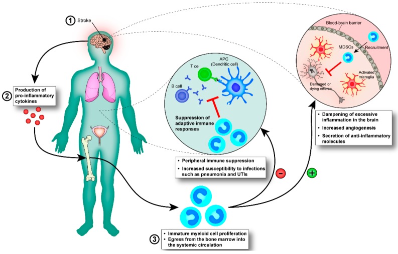
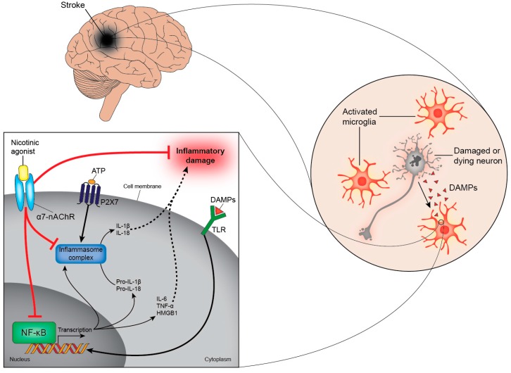
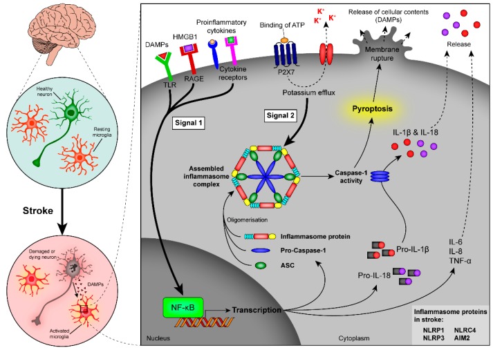
Stroke is one of the leading causes of death and long-term disability, with limited treatment options available. Inflammation contributes to damage tissue in the central nervous system across a broad range of neuropathologies, including Alzheimer's disease, pain, Schizophrenia, and stroke. While the immune system plays an important role in contributing to brain damage produced by ischemia, the damaged brain, in turn, can exert a powerful immune-suppressive effect that promotes infections and threatens the survival of stroke patients. Recently the cholinergic anti-inflammatory pathway, in particular its modulation using α7-nicotinic acetylcholine receptor (α7-nAChR) ligands, has shown potential as a strategy to dampen the inflammatory response and facilitate functional recovery in stroke patients. Here we discuss the current literature on stroke-induced inflammation and the effects of α7-nAChR modulators on innate immune cells.

Keywords: immune response; inflammation; myeloid cells; nicotinic; nicotinic acetylcholine receptor agonist; stroke.


Figures

Figure 1
Figure 1
Immature myeloid cells mediate immune-suppression post-stroke. (1) Stroke is followed by (2) the production of pro-inflammatory cytokines, which reach the bone marrow via the blood-stream (3) where they stimulate the expansion of immature myeloid cells and partially block their maturation into granulocytes, macrophages, and DCs. The immature myeloid cells (also known as MDSCs) migrate along a chemokine/cytokine gradient to secondary lymphoid organs and to the infarction area where they exert anti-inflammatory effects on other cell populations. (−) Immature myeloid cells are able to suppress innate and adaptive immune responses in the periphery, exposing stroke patients to an increased risk of acquiring infections, such as pneumonia and urinary tract infections (UTIs), which impede recovery and can potentially be life-threatening. (+) We hypothesise that immature myeloid cells travelling from the bone marrow to the brain exert protective effects there. Immature myeloid cells are highly phagocytic and would help clearing the infarct area of dead cells, and they are known to dampen inflammation through the secretion of cytokines that skew innate immune cells towards an anti-inflammatory phenotype. Furthermore, they are able to induce angiogenesis through the secretion of MMPs, which facilitates re-vascularisation. All of these effects would be beneficial, acting to limit excessive inflammation and promote healing. MMPs, matrix metalloproteinases.
Figure 2
Figure 2
Activation of α7-nAChRs reduces inflammation. Damaged and dying cells in the brain release DAMPs after a stroke. DAMPs can bind to several receptors, including TLRs, cytokine receptors, and the receptor for advanced glycation end products (RAGE). Ligation of these receptors activates NF-κB to translocate to the nucleus (bold arrow) where it induces the transcription of genes that code for pro-inflammatory cytokines, such as IL-6, IL-8, TNF-α and HMGB1. In addition, activation of NF-κB stimulates the production of the immature forms of IL-1β and IL-18 (pro-IL-1β and pro-IL-18, respectively). These biologically-inactive cytokines are cleaved to their active form by inflammasomes and are subsequently secreted from the cell (dashed arrows). These intracellular multi-protein complexes assemble in response to NF-κB stimulation and require a second stimulus (such as ATP) to become activated. α7-nAChR agonists interfere with these pathways and prevent the secretion of pro-inflammatory cytokines by inhibiting NF-κB and inflammasome activation (bar-headed red lines). α7-nAChRs, α7-nicotinic acetylcholine receptor; DAMPs, damage-associated molecular patterns; TLRs, toll-like receptors; NF-κB, nuclear factor kappa B; IL, interleukin; TNF-α, tumour necrosis factor-α; HMGB1, high-mobility group box 1; ATP, adenosine triphosphate.
Figure 3
Figure 3
Induction of inflammasome activity and pyroptosis after stroke. Signalling through PRRs results in NF-κB activation, which is associated with up-regulated transcription of inflammasome components (inflammasome proteins, pro-caspase-1, and ASC) and the immature cytokines pro-IL-1β and pro-IL-18, as well as an increased production of pro-inflammatory cytokines, such as IL-6, IL-8, and TNF-α (signal 1, bold arrow). A second signal (signal 2, bold arrow) leads to oligomerisation of the inflammasome components and activates the inflammasome complex. Signal 2 can, for example, occur through binding of ATP to its P2X7 receptor. As a result of inflammasome assembly and activation, caspase-1 gets activated and cleaves the inactive cytokines pro-IL-1β and pro-IL-18 to their active form, which are released from the cell (dashed arrows). Caspase-1 is also an important mediator of pyroptosis, a highly-inflammatory form of cell death, which results in the swelling and bursting of cells with the subsequent release of cell content into the extracellular space. PRRs, pattern recognition receptors; NF-κB, nuclear factor kappa B; ASC, apoptosis-associated speck-like protein containing a CARD; IL, interleukin; tumour necrosis factor-α; ATP, adenosine triphosphate.

References

    1. Dirnagl U., Iadecola C., Moskowitz M.A. Pathobiology of ischaemic stroke: An integrated view. Trends Neurosci. 1999;22:391–397. doi: 10.1016/S0166-2236(99)01401-0. - DOI - PubMed
    1. Martinon F. Detection of immune danger signals by nalp3. J. Leukoc. Biol. 2008;83:507–511. doi: 10.1189/jlb.0607362. - DOI - PubMed
    1. Martin W.J., Walton M., Harper J. Resident macrophages initiating and driving inflammation in a monosodium urate monohydrate crystal-induced murine peritoneal model of acute gout. Arthritis Rheumatol. 2009;60:281–289. doi: 10.1002/art.24185. - DOI - PubMed
    1. Chamorro A., Urra X., Planas A.M. Infection after acute ischemic stroke: A manifestation of brain-induced immunodepression. Stroke. 2007;38:1097–1103. doi: 10.1161/01.STR.0000258346.68966.9d. - DOI - PubMed
    1. Liesz A., Dalpke A., Mracsko E., Antoine D.J., Roth S., Zhou W., Yang H., Na S.Y., Akhisaroglu M., Fleming T., et al. Damp signaling is a key pathway inducing immune modulation after brain injury. J. Neurosci. 2015;35:583–598. doi: 10.1523/JNEUROSCI.2439-14.2015. - DOI - PMC - PubMed

Publication types

Substances

LinkOut - more resources