. 2015 Dec 22:5:18599.
doi: 10.1038/srep18599.

Secreted Factors from Human Vestibular Schwannomas Can Cause Cochlear Damage

Affiliations

Secreted Factors from Human Vestibular Schwannomas Can Cause Cochlear Damage

Sonam Dilwali et al. Sci Rep. .

Abstract

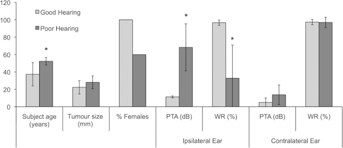
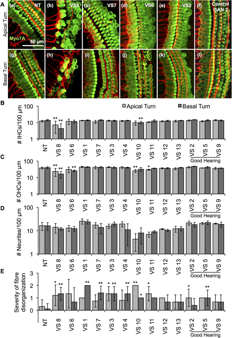
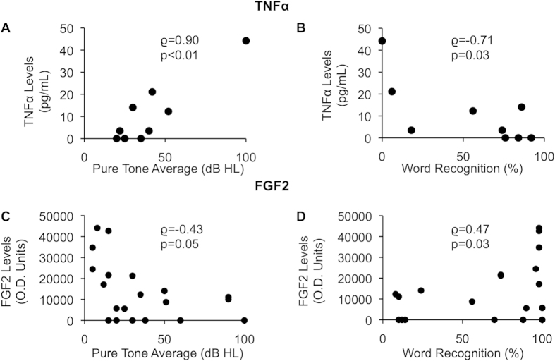
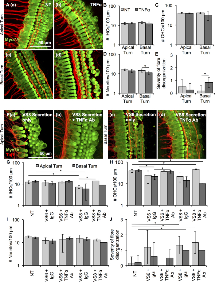
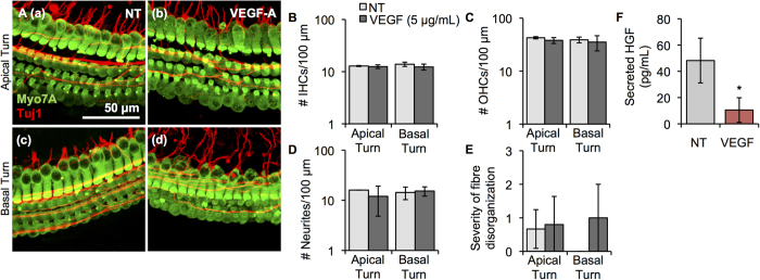
Vestibular schwannomas (VSs) are the most common tumours of the cerebellopontine angle. Ninety-five percent of people with VS present with sensorineural hearing loss (SNHL); the mechanism of this SNHL is currently unknown. To establish the first model to study the role of VS-secreted factors in causing SNHL, murine cochlear explant cultures were treated with human tumour secretions from thirteen different unilateral, sporadic VSs of subjects demonstrating varied degrees of ipsilateral SNHL. The extent of cochlear explant damage due to secretion application roughly correlated with the subjects' degree of SNHL. Secretions from tumours associated with most substantial SNHL resulted in most significant hair cell loss and neuronal fibre disorganization. Secretions from VSs associated with good hearing or from healthy human nerves led to either no effect or solely fibre disorganization. Our results are the first to demonstrate that secreted factors from VSs can lead to cochlear damage. Further, we identified tumour necrosis factor alpha (TNFα) as an ototoxic molecule and fibroblast growth factor 2 (FGF2) as an otoprotective molecule in VS secretions. Antibody-mediated TNFα neutralization in VS secretions partially prevented hair cell loss due to the secretions. Taken together, we have identified a new mechanism responsible for SNHL due to VSs.


Figures

Figure 1
Figure 1. Patient demographics for VS secretions applied to cochlear explants.
Average subject age in years, tumour size (mm, largest transverse dimension), % females, pure tone average (PTA, dB), and word recognition score (WR, %) are given for the ipsilateral and contralateral ears to VS. Data are segregated into groups of VS associated with good hearing (PTA < 30 dB and WR > 70%, n = 3 being VS2, VS5 and VS9) and poor hearing (n = 10). * = p < 0.05.
Figure 2
Figure 2. Application of human VS secretions onto murine cochlear explant cultures leads to hair cell and neurite loss.
(A). Representative images for cochlear explants receiving (a) no treatment (NT, n = 28 different explants), incubated with (b) VS8 (n = 5 different explants), (c) VS7 (n = 4 different explants), (d) VS6 (n = 3 different explants), (e) VS2 (n = 5 different explants) and (f) control GAN2 (n = 3 different explants) secretions are shown for the apical turns, and (g) NT (n = 26 different explants), (h) VS8 (n = 6 different explants), (i) VS7 (n = 5 different explants), (j) VS6 (n = 3 different explants), (k) VS2 (n = 5 different explants) and (l) control GAN2 (n = 4 different explants) secretions for the basal turn. Myo7A (green) marks hair cells and Tuj1 (red) marks neurites. Scale Bar = 50 μm applies to all images. (B). Number of inner hair cells (IHCs), (C). outer hair cells (OHCs), (D). neurites, and (E). severity of fibre disorganization are shown for a 100 μm length within the apex (light grey columns) and basal turn (dark grey columns) cochlear explants treated with NT and secretions from 13 different tumours. *p < 0.05, **p < 0.01. Quantified data after treatment with control GAN secretions are shown in Supplementary Figure 1.
Figure 3
Figure 3. Correlation of VS-secreted TNFα and FGF2 with SNHL due to VS.
(A). Correlation of pure tone average (dB) and (B). word recognition score (%) of ipsilateral ear to the VS, and measured secreted TNFα levels from VSs (n = 9). (C). Correlation of pure tone average (dB) and (D). word recognition score (%) of ipsilateral ear to the VS and measured secreted FGF2 levels from VSs (n = 21). p-values are shown, rho represents Spearman’s rank correlation coefficient.
Figure 4
Figure 4. TNFα application onto cochlear explants induced mild damage.
(A). Representative images for cochlear explants receiving no treatment (NT, a, c, n = 6 different explants) or incubated with TNFα (b, d, n = 4–6 different explants) are shown for the apical (a, b) and basal (c, d) turn. Myo7A (green) marks hair cells and Tuj1 (red) marks neurites. Scale Bar = 50 μm applies to all images. (B). Number of inner hair cells (IHCs), (C). number of outer hair cells (OHCs), (D). number of neurites, (E). severity of fibre disorganization are shown for a 100 μm length within the apical and basal turn explants for NT (light grey columns) and TNFα-treated (dark grey columns). *p < 0.05. (F). Representative images for cochlear explants receiving VS secretions only (a, c, n = 2 different tumours) or incubated with VS secretions with TNFα antibody (b, d, n = 2 different tumours) are shown for the apical (a, b) and basal (c, d) turn. Myo7A (green) marks hair cells and Tuj1 (red) marks neurites. Scale Bar = 50 μm applies to all images. (G). Number of inner hair cells (IHCs), (H). number of outer hair cells (OHCs), (I). number of neurites, (J). severity of fibre disorganization are shown for a 100 μm length within the apical and basal turn explants treated with VS secretions only (light grey columns) and VS secretions with TNFα antibody (dark grey columns). *p < 0.05.
Figure 5
Figure 5. VEGF-A application onto cochlear explants did not induce substantial cellular damage but did significantly decrease HGF secretion.
(A). Representative images for cochlear explants receiving no treatment (NT, a, c, n = 3–4 different explants) or incubated with VEGF-A (b, d, n = 3–5 different explants) are shown for the apical (a, b) and basal (c, d) turn. Myo7A (green) marks hair cells and Tuj1 (red) marks neurites. Scale Bar = 50 μm applies to all images. (B). Number of inner hair cells (IHCs), (C). number of outer hair cells (OHCs), (D). number of neurites, (E). severity of fibre disorganization are shown for a 100 μm length within the apical and basal turn explants for NT (light grey columns) and VEGF-A-treated (dark grey columns) (F). Secreted murine HGF levels in cochlear explants post-VEGF treatment. *p < 0.05.

Similar articles

Cited by

References

    1. Thakur J. D. et al. An update on unilateral sporadic small vestibular schwannoma. Neurosurg. Focus 33, E1, doi: 10.3171/2012.6.FOCUS12144 (2012). - DOI - PubMed
    1. Mahaley M. S. Jr., Mettlin C., Natarajan N., Laws E. R. Jr. & Peace B. B. Analysis of patterns of care of brain tumor patients in the United States: a study of the Brain Tumor Section of the AANS and the CNS and the Commission on Cancer of the ACS. Clin. Neurosurg. 36, 347–352 (1990). - PubMed
    1. Plotkin S. R. et al. Bevacizumab for progressive vestibular schwannoma in neurofibromatosis type 2: a retrospective review of 31 patients. Otol. Neurotol. 33, 1046–1052, doi: 10.1097/MAO.0b013e31825e73f5 (2012). - DOI - PubMed
    1. Aronzon A., Ruckenstein M. J. & Bigelow D. C. The efficacy of corticosteroids in restoring hearing in patients undergoing conservative management of acoustic neuromas. Otol. Neurotol. 24, 465–468 (2003). - PubMed
    1. Nadol J. B. Jr., Diamond P. F. & Thornton A. R. Correlation of hearing loss and radiologic dimensions of vestibular schwannomas (acoustic Neuromas). Am. J. Otol. 17, 312–316 (1996). - PubMed

Publication types

MeSH terms

Substances