

Adaptive evolution in the toxicity of a spider's venom enzymes
- PMID: 26690570
- PMCID: PMC4687385
- DOI: 10.1186/s12862-015-0561-4
Adaptive evolution in the toxicity of a spider's venom enzymes
Erratum in
-
Erratum: Adaptive evolution in the toxicity of a spider's venom enzymes.BMC Evol Biol. 2016 Mar 7;16:58. doi: 10.1186/s12862-016-0623-2. BMC Evol Biol. 2016. PMID: 26951516 Free PMC article. No abstract available.
Abstract
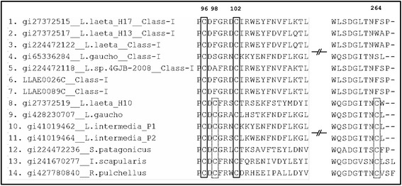
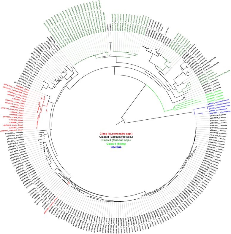
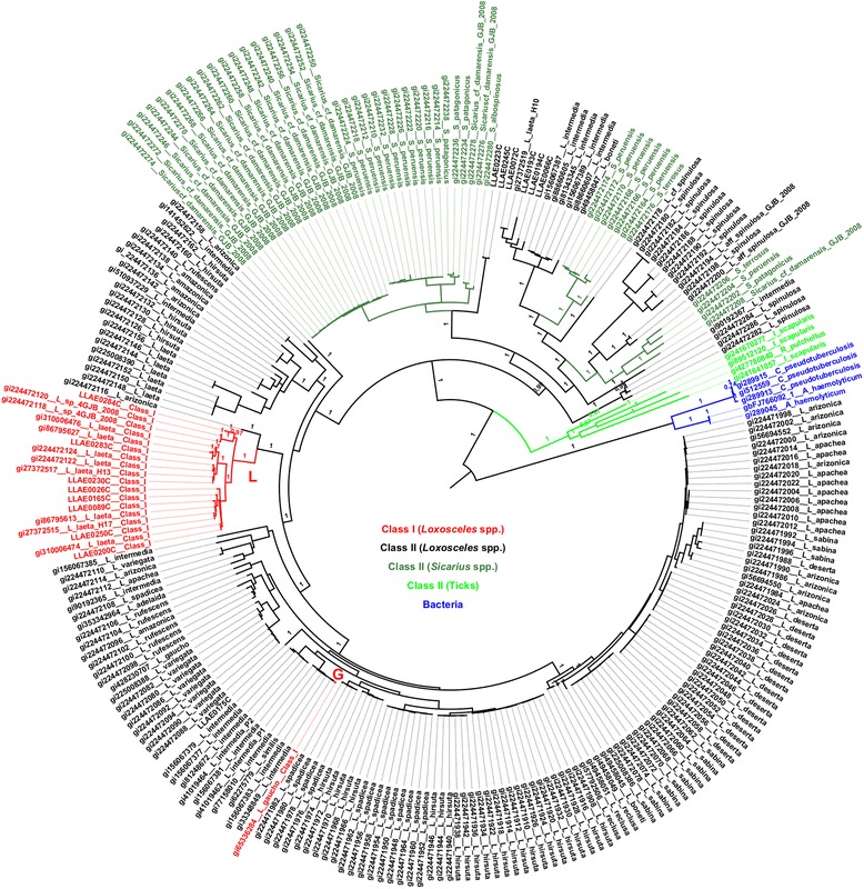
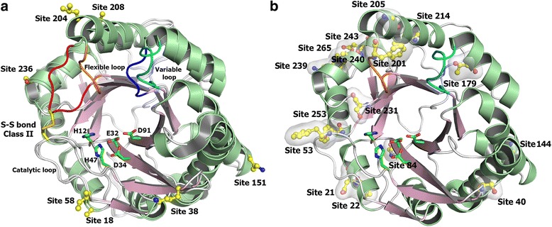
Background: Sphingomyelinase D is the main toxin present in the venom of Loxosceles spiders. Several isoforms present in these venoms can be structurally classified in two groups. Class I Sphingomyelinase D contains a single disulphide bridge and variable loop. Class II Sphingomyelinase D presents an additional intrachain disulphide bridge that links a flexible loop with a catalytic loop. These classes exhibit differences in their toxic potential. In this paper we address the distribution of the structural classes of SMase D within and among species of spiders and also their evolutionary origin by means of phylogenetic analyses. We also conducted tests to assess the action of natural selection in their evolution combined to structural modelling of the affected sites.
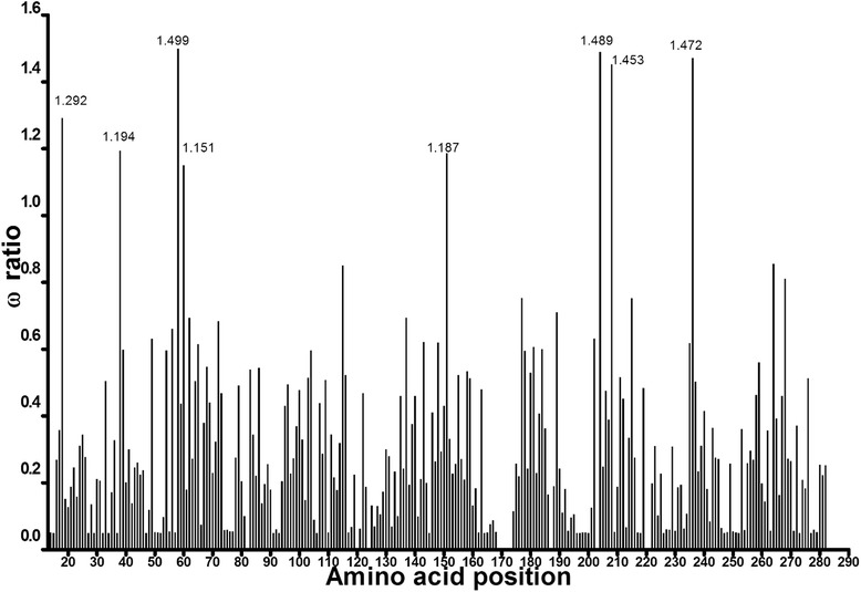
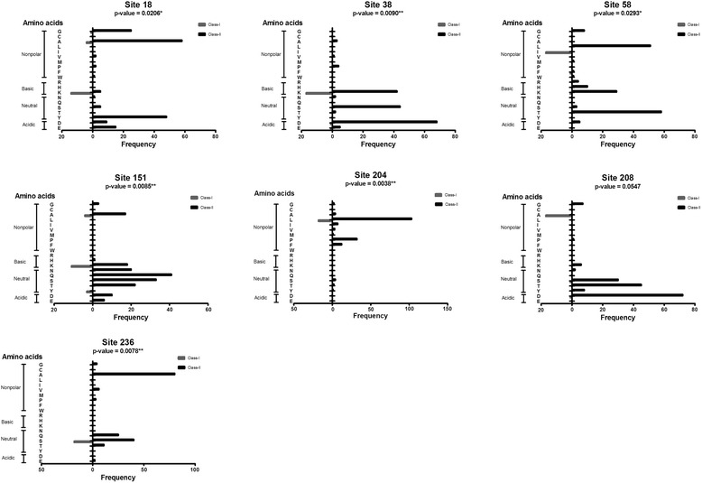
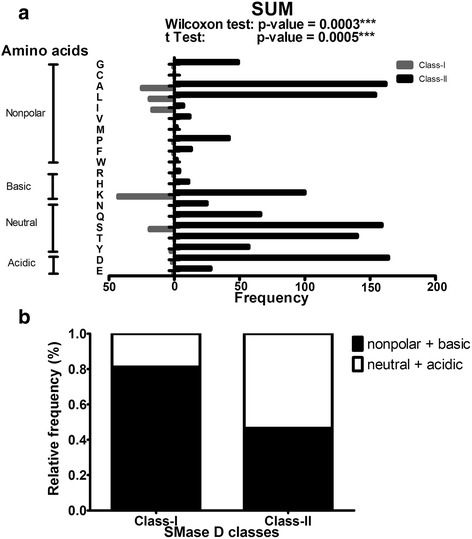
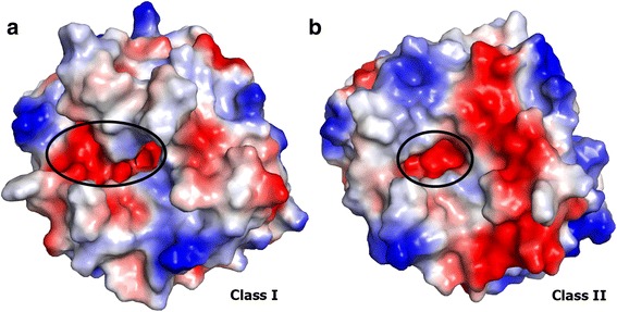
Results: The majority of the Class I enzymes belong to the same clade, which indicates a recent evolution from a single common ancestor. Positively selected sites are located on the catalytic interface, which contributes to a distinct surface charge distribution between the classes. Sites that may prevent the formation of an additional bridge were found in Class I enzymes.
Conclusions: The evolution of Sphingomyelinase D has been driven by natural selection toward an increase in noxiousness, and this might help explain the toxic variation between classes.
Figures

Similar articles
-
Sphingomyelinase D Activity in Sicarius tropicus Venom: Toxic Potential and Clues to the Evolution of SMases D in the Sicariidae Family.Toxins (Basel). 2021 Apr 1;13(4):256. doi: 10.3390/toxins13040256. Toxins (Basel). 2021. PMID: 33916208 Free PMC article.
-
Sphingomyelinase D from venoms of Loxosceles spiders: evolutionary insights from cDNA sequences and gene structure.Toxicon. 2005 Apr;45(5):547-60. doi: 10.1016/j.toxicon.2004.11.011. Toxicon. 2005. PMID: 15777950
-
The phylogenetic distribution of sphingomyelinase D activity in venoms of Haplogyne spiders.Comp Biochem Physiol B Biochem Mol Biol. 2003 May;135(1):25-33. doi: 10.1016/s1096-4959(03)00045-9. Comp Biochem Physiol B Biochem Mol Biol. 2003. PMID: 12781970
-
Biotechnological applications of brown spider (Loxosceles genus) venom toxins.Biotechnol Adv. 2008 May-Jun;26(3):210-8. doi: 10.1016/j.biotechadv.2007.12.003. Epub 2007 Dec 23. Biotechnol Adv. 2008. PMID: 18207690 Review.
-
Spider's venom phospholipases D: A structural review.Int J Biol Macromol. 2018 Feb;107(Pt A):1054-1065. doi: 10.1016/j.ijbiomac.2017.09.081. Epub 2017 Sep 23. Int J Biol Macromol. 2018. PMID: 28951301 Review.
Cited by
-
Unveiling the Protein Components of the Secretory-Venom Gland and Venom of the Scorpion Centruroides possanii (Buthidae) through Omic Technologies.Toxins (Basel). 2023 Aug 9;15(8):498. doi: 10.3390/toxins15080498. Toxins (Basel). 2023. PMID: 37624255 Free PMC article.
-
Digestive enzymes and sphingomyelinase D in spiders without venom (Uloboridae).Sci Rep. 2023 Feb 15;13(1):2661. doi: 10.1038/s41598-023-29828-x. Sci Rep. 2023. PMID: 36792649 Free PMC article.
-
Differential Cellular Responses to Class I and II Sphingomyelinase D: Unraveling the Mechanisms of Loxosceles Venom-Induced Dermonecrosis and Potential Therapeutic Targets.Int J Mol Sci. 2025 Mar 26;26(7):3012. doi: 10.3390/ijms26073012. Int J Mol Sci. 2025. PMID: 40243660 Free PMC article.
-
Sphingomyelinase D Activity in Sicarius tropicus Venom: Toxic Potential and Clues to the Evolution of SMases D in the Sicariidae Family.Toxins (Basel). 2021 Apr 1;13(4):256. doi: 10.3390/toxins13040256. Toxins (Basel). 2021. PMID: 33916208 Free PMC article.
-
The Dual Prey-Inactivation Strategy of Spiders-In-Depth Venomic Analysis of Cupiennius salei.Toxins (Basel). 2019 Mar 19;11(3):167. doi: 10.3390/toxins11030167. Toxins (Basel). 2019. PMID: 30893800 Free PMC article.
References
-
- Tambourgi DV, Magnoli FC, van den Berg CW, Morgan BP, de Araujo PS, Alves EW, et al. Sphingomyelinases in the venom of the spider Loxosceles intermedia are responsible for both dermonecrosis and complement-dependent hemolysis. Biochem Biophys Res Commun. 1998;251:366–373. doi: 10.1006/bbrc.1998.9474. - DOI - PubMed
Publication types
MeSH terms
Substances
LinkOut - more resources
Full Text Sources
Other Literature Sources
