. 2016 Feb 15:422:160-171.
doi: 10.1016/j.mce.2015.11.033. Epub 2015 Dec 13.

GPER1-mediated IGFBP-1 induction modulates IGF-1-dependent signaling in tamoxifen-treated breast cancer cells

Affiliations

GPER1-mediated IGFBP-1 induction modulates IGF-1-dependent signaling in tamoxifen-treated breast cancer cells

Ali Vaziri-Gohar et al. Mol Cell Endocrinol. .

Abstract

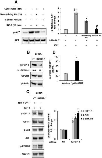
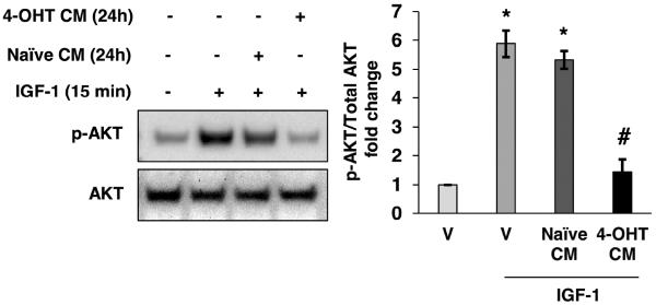
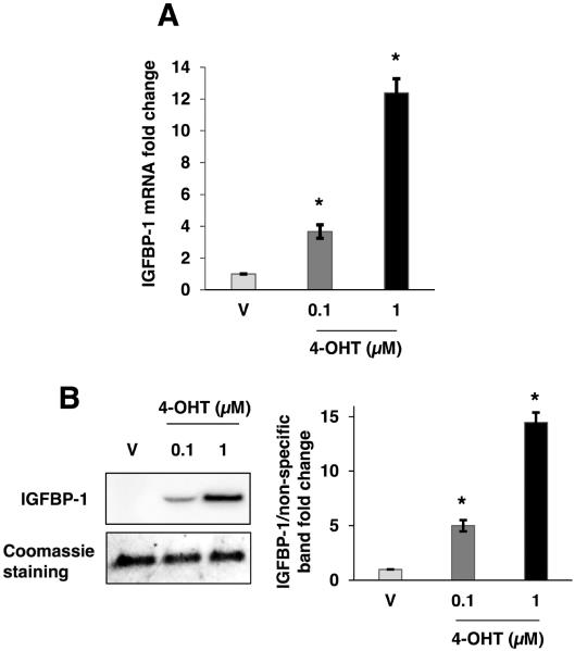
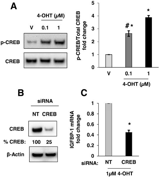
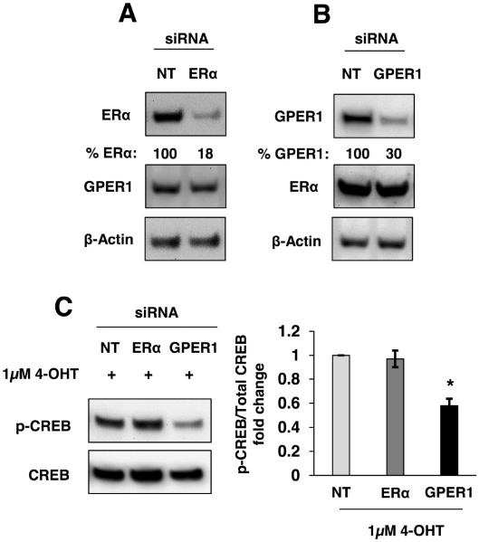
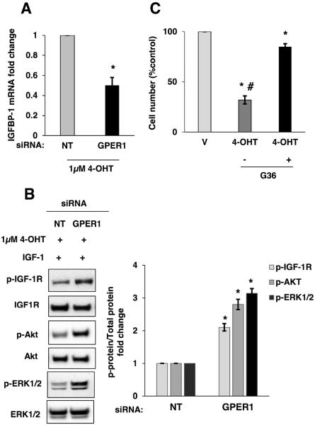
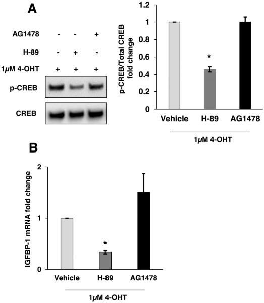
Tamoxifen, a selective estrogen receptor modulator, is a commonly prescribed adjuvant therapy for estrogen receptor-α (ERα)-positive breast cancer patients. To determine if extracellular factors contribute to the modulation of IGF-1 signaling after tamoxifen treatment, MCF-7 cells were treated with IGF-1 in conditioned medium (CM) obtained from 4-OHT-treated MCF-7 cells and the accumulation of phospho-Akt (S473) was measured. CM inhibited IGF-1-dependent cell signaling and suggesting the involvement of extracellular factors (ie. IGFBPs). A significant increase in IGFBP-1 mRNA and extracellular IGFBP-1 protein was observed in 4-OHT-treated MCF-7 cells. Knockdown experiments demonstrated that both GPER1 and CREB mediate IGFBP-1 induction. Furthermore, experiments showed that 4-OHT-dependent IGFBP-1 transcription is downstream of GPER1-activation in breast cancer cells. Additionally, neutralization and knockdown experiments demonstrated a role for IGFBP-1 in the observed inhibition of IGF-1 signaling. These results suggested that 4-OHT inhibits IGF-1 signaling via GPER1 and CREB mediated extracellular IGFBP-1 accumulation in breast cancer cells.

Keywords: Breast cancer; GPER1; IGF-1; IGFBP-1; Tamoxifen.


Figures

Fig. 1
Fig. 1
4-OHT-induced extracellular factor inhibits IGF-1-stimulated AKT phosphorylation. Conditioned medium (CM) from vehicle-treated MCF-7 cells (24 hours) was replaced with conditioned medium from 4-OHT-treated cells prior to IGF-1 stimulation (50 ng/mL) for 15 min. Total AKT and phospho-AKT were measured by immunoblot. *, p<0.05 compared to vehicle (V) control, and #, p<0.05 compared to IGF-1 treated sample. The results are representative of three independent experiments, and the error bars are SEM.
Fig. 2
Fig. 2
4-OHT induced IGFBP-1 transcription and extracellular accumulation in MCF-7 cells. (A) Relative IGFBP-1 gene transcription determined by quantitative real-time PCR after 24 hours of treatment with vehicle (V), 100 nM 4-OHT, or 1μM 4-OHT. Relative transcription was normalized to the RPL30 transcript. (B) Immunoblot analysis of concentrated MCF-7 cell culture medium after 24 hours of treatment with vehicle (V), 100 nM 4-OHT, or 1μM 4-OHT. Results are representative of at least three independent experiments. *, p<0.05 compared to vehicle control, and the error bars are SEM.
Fig. 3
Fig. 3
CREB was phosphorylated after treatment with 4-OHT and CREB knockdown partially inhibited 4-OHT-induced IGFBP-1 transcription in MCF-7 cells. (A) Immunoblot analysis of phospho-CREB (S133) after 2 hours of treatment with 4-OHT using the indicated doses. (B) Immunoblot analysis of CREB expression after transfection of CREB-targeting siRNA. (C) Relative IGFBP-1 gene transcription determined by quantitative real-time PCR in CREB knockdown and control cells (NT) after 24 hours of treatment with 4-OHT. *, p<0.05 compared to control, and #, p<0.05 compared to 4-OHT (1μM) treated sample. The results are representative of three independent experiments, and the error bars are SEM.
Fig. 4
Fig. 4
GPER1-mediated CREB phosphorylation in MCF-7 cells. (A, B) The total ERα and GPER1 protein levels were reduced using siRNA knockdown. (C) Immunoblot analysis of CREB phosphorylation after 2 hours of treatment with 1μM 4-OHT subsequent to ERα or GPER1 knockdown compared to non-targeting siRNA (NT) cells. *, p<0.05 compared to control. The results are representative of three independent experiments, and the error bars are SEM.
Fig. 5
Fig. 5
4-OHT-induced IGFBP-1 transcription and inhibition of IGF-1 stimulation is decreased after GPER1 knockdown in MCF-7 cells. (A) Relative IGFBP-1 gene transcription determined by quantitative real-time PCR in GPER1 knockdown and non-targeting control cells (NT) after 24 hours of treatment with vehicle or 1μM 4-OHT. (B) Immunoblot analysis p-IGF-1R, p-AKT and p-ERK1/2 after IGF-1 stimulation in control and GPER1 knockdown MCF-7 cells. (C) MCF-7 cell viability after treatment with 1μM 4-OHT with and without 1μM G36 treatment. *, p<0.05 compared to vehicle and #, p<0.05 compared to G36- treated sample. The results are representative of three independent experiments, and the error bars are SEM.
Fig. 6
Fig. 6
4-OHT-induced IGFBP-1 transcription is mediated by PKA in MCF-7 cells. (A) Immunoblot analysis of CREB phosphorylation in PKA-selective inhibitor (H-89) and EGFR-selective inhibitor (AG-1478) followed by 4-OHT treatment for 2h. (B) Relative IGFBP-1 gene transcription determined by quantitative real-time PCR in 20 μM H-89 and 20 μM AG1478 treated MCF-7 cells after 24 hours of treatment with 1μM 4-OHT. *, p<0.05 compared to control. The results are representative of three independent experiments, and the error bars are SEM.
Fig. 7
Fig. 7
Antibody-mediated IGFBP-1 neutralization or IGFBP-1 knockdown reverses the inhibition of IGF-1 stimulation in 4-OHT-treated MCF-7 cells. (A) Two hours prior to IGF-1 stimulation (15 minutes), control IgG or IGFBP-1 neutralizing antibody (10 ng/mL each) were added to MCF-7 cells treated with vehicle (V) or 4-OHT-treated for 24 hours. Total AKT and phospho-AKT levels were determined by immunoblot. (B) siRNA mediated reduction of IGFBP-1 protein expression. (C) After transfection with IGFBP-1 siRNAs or non-targeting control (NT), MCF-7 cells were treated with 1μM 4-OHT for 24 hours followed by 50 ng/mL IGF-1 stimulation for 15 minutes followed by the immunoblot analysis of p-IGF-1R (Y1135/1136), p-AKT (S473) and p-ERK1/2. (D) Extracellular IGFBP-1 protein concentration determined by ELISA. *, p<0.05 compared to vehicle, and #, p<0.05 compared to IGFBP-1 blocking Ab-treated. The results are representative of three independent experiments, and the error bars are SEM.

Similar articles

Cited by

References

    1. Ariazi EA, Brailoiu E, Yerrum S, Shupp HA, Slifker MJ, Cunliffe HE, Black MA, Donato AL, Arterburn JB, Oprea TI, Prossnitz ER, Dun NJ, Jordan VC. The G Protein-Coupled Receptor GPR30 Inhibits Proliferation of Estrogen Receptor-Positive Breast Cancer Cells. Cancer Res. 2010;70:1184–1194. - PMC - PubMed
    1. Arteaga CL, Kitten LJ, Coronado EB, Jacobs S, Kull FC, Allred DC, Osborne CK. Blockade of the type-I somatomedin receptor inhibits growth of human-breast cancer-cells in athymic mice. J. Clin. Invest. 1989;84:1418–1423. - PMC - PubMed
    1. Arteaga CL. Interference of the IGF system as a strategy to inhibit breast-cancer growth. Breast Cancer Res. Treat. 1992;22:101–106. - PubMed
    1. Baxter RC. Insulin-like growth factor binding protein-3 (IGFBP-3): Novel ligands mediate unexpected functions. J. Cell Commun. Signal. 2013;7:179–189. - PMC - PubMed
    1. Becker MA, Ibrahim YH, Cui X, Lee AV, Yee D. The IGF Pathway Regulates ER alpha through a S6K1-Dependent Mechanism in Breast Cancer Cells. Mol. Endocrinol. 2011;25:516–528. - PMC - PubMed

Publication types

MeSH terms

Substances