

Longitudinal changes in resting-state fMRI from age 5 to age 6years covary with language development
- PMID: 26690809
- PMCID: PMC4767215
- DOI: 10.1016/j.neuroimage.2015.12.008
Longitudinal changes in resting-state fMRI from age 5 to age 6years covary with language development
Abstract
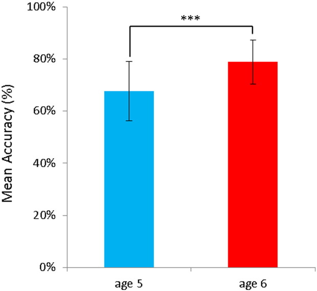
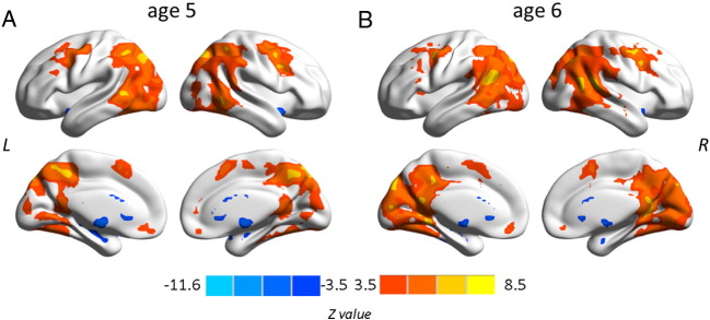
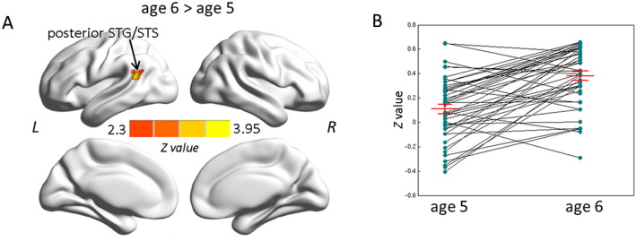
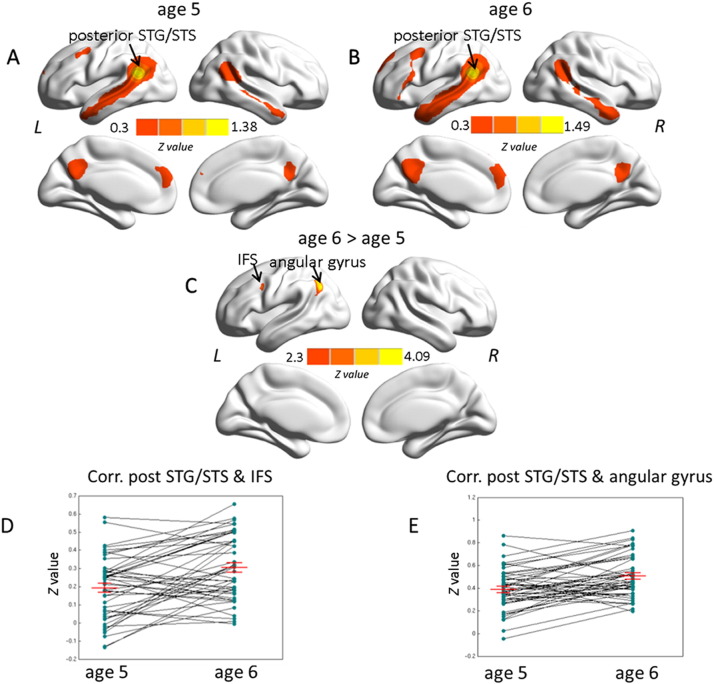
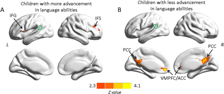
Resting-state functional magnetic resonance imaging is a powerful technique to study the whole-brain neural connectivity that underlies cognitive systems. The present study aimed to define the changes in neural connectivity in their relation to language development. Longitudinal resting-state functional data were acquired from a cohort of preschool children at age 5 and one year later, and changes in functional connectivity were correlated with language performance in sentence comprehension. For this, degree centrality, a voxel-based network measure, was used to assess age-related differences in connectivity at the whole-brain level. Increases in connectivity with age were found selectively in a cluster within the left posterior superior temporal gyrus and sulcus (STG/STS). In order to further specify the connection changes, a secondary seed-based functional connectivity analysis on this very cluster was performed. The correlations between resting-state functional connectivity (RSFC) and language performance revealed developmental effects with age and, importantly, also dependent on the advancement in sentence comprehension ability over time. In children with greater advancement in language abilities, the behavioral improvement was positively correlated with RSFC increase between left posterior STG/STS and other regions of the language network, i.e., left and right inferior frontal cortex. The age-related changes observed in this study provide evidence for alterations in the language network as language develops and demonstrates the viability of this approach for the investigation of normal and aberrant language development.
Keywords: Frontal-to-temporal connection; Intrinsic connectivity; Language development; Preschool children; Resting-state fMRI.
Copyright © 2015 The Authors. Published by Elsevier Inc. All rights reserved.
Figures
Similar articles
-
Development of the Intrinsic Language Network in Preschool Children from Ages 3 to 5 Years.PLoS One. 2016 Nov 3;11(11):e0165802. doi: 10.1371/journal.pone.0165802. eCollection 2016. PLoS One. 2016. PMID: 27812160 Free PMC article.
-
Development of a selective left-hemispheric fronto-temporal network for processing syntactic complexity in language comprehension.Neuropsychologia. 2016 Mar;83:274-282. doi: 10.1016/j.neuropsychologia.2015.09.003. Epub 2015 Sep 6. Neuropsychologia. 2016. PMID: 26352468 Free PMC article.
-
Longitudinal Study of the Emerging Functional Connectivity Asymmetry of Primary Language Regions during Infancy.J Neurosci. 2016 Oct 19;36(42):10883-10892. doi: 10.1523/JNEUROSCI.3980-15.2016. J Neurosci. 2016. PMID: 27798142 Free PMC article.
-
Biophysical and neural basis of resting state functional connectivity: Evidence from non-human primates.Magn Reson Imaging. 2017 Jun;39:71-81. doi: 10.1016/j.mri.2017.01.020. Epub 2017 Feb 2. Magn Reson Imaging. 2017. PMID: 28161319 Free PMC article. Review.
-
Resting-State Functional Connectivity: Signal Origins and Analytic Methods.Neuroimaging Clin N Am. 2020 Feb;30(1):15-23. doi: 10.1016/j.nic.2019.09.012. Neuroimaging Clin N Am. 2020. PMID: 31759568 Review.
Cited by
-
Effects of ambient fine particulates, nitrogen dioxide, and ozone on maturation of functional brain networks across early adolescence.Environ Int. 2023 Jul;177:108001. doi: 10.1016/j.envint.2023.108001. Epub 2023 Jun 1. Environ Int. 2023. PMID: 37307604 Free PMC article.
-
Atypical functional connectivity of temporal cortex with precuneus and visual regions may be an early-age signature of ASD.Mol Autism. 2023 Mar 10;14(1):11. doi: 10.1186/s13229-023-00543-8. Mol Autism. 2023. PMID: 36899425 Free PMC article.
-
Neuroanatomical basis of language ability in an autism subgroup with moderate language deficits.Eur Child Adolesc Psychiatry. 2025 Jun;34(6):1895-1904. doi: 10.1007/s00787-024-02605-5. Epub 2024 Nov 8. Eur Child Adolesc Psychiatry. 2025. PMID: 39514012
-
Language Network Function in Young Children Born Very Preterm.Front Hum Neurosci. 2018 Dec 20;12:512. doi: 10.3389/fnhum.2018.00512. eCollection 2018. Front Hum Neurosci. 2018. PMID: 30618688 Free PMC article.
-
Sources of Heterogeneity in Functional Connectivity During English Word Processing in Bilingual and Monolingual Children.Neurobiol Lang (Camb). 2023 Apr 11;4(2):198-220. doi: 10.1162/nol_a_00092. eCollection 2023. Neurobiol Lang (Camb). 2023. PMID: 37229508 Free PMC article.
References
Publication types
MeSH terms
LinkOut - more resources
Full Text Sources
Other Literature Sources
