. 2016 Mar;238(4):584-97.
doi: 10.1002/path.4682.

Decreased FOXJ1 expression and its ciliogenesis programme in aggressive ependymoma and choroid plexus tumours

Affiliations

Decreased FOXJ1 expression and its ciliogenesis programme in aggressive ependymoma and choroid plexus tumours

Malak S Abedalthagafi et al. J Pathol. 2016 Mar.

Abstract

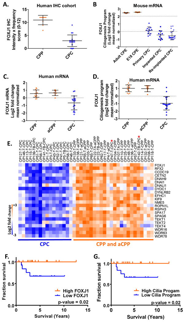
Well-differentiated human cancers share transcriptional programmes with the normal tissue counterparts from which they arise. These programmes broadly influence cell behaviour and function and are integral modulators of malignancy. Here, we show that the master regulator of motile ciliogenesis, FOXJ1, is highly expressed in cells along the ventricular surface of the human brain. Strong expression is present in cells of the ependyma and the choroid plexus as well as in a subset of cells residing in the subventricular zone. Expression of FOXJ1 and its transcriptional programme is maintained in many well-differentiated human tumours that arise along the ventricle, including low-grade ependymal tumours and choroid plexus papillomas. Anaplastic ependymomas as well as choroid plexus carcinomas show decreased FOXJ1 expression and its associated ciliogenesis programme genes. In ependymomas and choroid plexus tumours, reduced expression of FOXJ1 and its ciliogenesis programme are markers of poor outcome and are therefore useful biomarkers for assessing these tumours. Transitions in ciliogenesis define distinct differentiation states in ependymal and choroid plexus tumours with important implications for patient care. Copyright © 2015 Pathological Society of Great Britain and Ireland. Published by John Wiley & Sons, Ltd.

Keywords: FOXJ1; choroid plexus; cilia; ciliogenesis; dedifferentiation; ependymoma; stem cells; subventricular zone.


Conflict of interest statement

Conflict of interest: No disclosures.

Figures

Figure 1
Figure 1
FOXJ1 and critical components of the ciliogenesis program are highly expressed in ependymoma. A. Heat map of normalized publicly-available expression profiling data in the Gene Expression Omnibus (GEO) of various brain tumours (n = 346) and normal brain tissues (n =10 samples). Columns represent the average log2 fold change from mean normalized data for each sample type (Table S1). High expression orange, low expression blue. Transcription factors that regulate ciliogenesis as well as genes that are major components of cilia are shown. Pilocytic astrocytoma (n = 41), ependymoma (n = 19), meningioma (n = 68), Central Nervous System (CNS) PNET (n = 22), high-grade astrocytoma (including glioblastoma; n = 84), medulloblastoma (n = 29), atypical teratoid/rhabdoid tumour (n = 21), primary CNS lymphoma (n = 34), pineoblastoma (n = 1), pediatric glioblastoma (n = 27), normal brain (n = 10). B. and C. Scatter plot of log2 fold change of the mean normalized data for the mRNA of the cilia transcriptional regulators FOXJ1 and RFX2. Each point represents the data from a single gene expression profile from the indicated tissue type (from Table S1).
Figure 2
Figure 2
FOXJ1 mRNA and protein are expressed in the ependymal lining and choroid plexus of the mouse and human brain. A. and B. FOXJ1 in situ hybridization in a P14 mouse brain (images from the Allen Developing Mouse Brain Atlas). Immunohistochemistry for FOXJ1 protein expression in ciliated cells lining C. the plicae of the human fallopian tube and D. the bronchial airways of the human lung. E. Immunohistochemistry for FOXJ1 protein expression in the ependymal line (strong) and the choroid plexus (weaker) of the mouse brain. F. H&E stained section and G. immunohistochemistry for FOXJ1 protein expression in the brain of a 76 year old woman. SVZ denotes the subventricular zone region. There are many scattered FOXJ1 immunoreactive cells in the subventricular zone of the adult human brain. H. RNAscope in situ hybridization for FOXJ1 mRNA in a serial section of the same brain. Scale bar sizes are indicated in each of the images.
Figure 3
Figure 3
FOXJ1 protein is expressed in various ependymoma subtypes. A. H&E stained section of a WHO grade II ependymoma and B. the corresponding FOXJ1 immunohistochemistry. C. H&E stained sections of a WHO grade I myxopapillary ependymoma and D. the corresponding FOXJ1 immunohistochemistry. E. FOXJ1 immunohistochemistry in a cellular ependymoma and F. FOXJ1 immunohistochemistry in a subependymoma. Scale bar sizes are indicated in each of the images.
Figure 4
Figure 4
Expression of FOXJ1 and the ciliogenesis program varies in ependymoma. A. Heat map of FOXJ1, RFX2 and the FOXJ1 ciliogenesis program (see Materials and Methods section for details of the program; Table S4) in ependymoma. The source data is from Witt et al. [24]. Locations of the tumours (supratentorial, posterior fossa and spinal) are listed (Table S6). B. Plot of FOXJ1 mRNA expression and the mean of the expression of FOXJ1 ciliogenesis program genes (average of mean fold change for the genes comprising this program). The source data is from Witt et al. [24]. Analysis of the correlation shows a Pearson r = 0.89. C. Plot of the FOXJ1 ciliogenesis program from data in Pajtler et al, [22] for posterior fossa ependymoma (PF-EPN-A or PF-EPN-B), spinal cord ependymoma (SP-EPN) or supratentorial ependymoma (ST) with RELA (ST-EPN-RELA) or YAP1 (ST-EPND-YAP) fusions (Table S7). D. H&E stained section and FOXJ1 immunohistochemistry from serial sections of a recurrent anaplastic ependymoma with dedifferentiated regions from the right parietal-occipital region of a 21 year old male. E. Plot of FOXJ1 immunohistochemistry score (intensity x frequency on a scale of 0–12) on a set of 97 ependymal tumours including 42 WHO grade II ependymoma, 30 WHO grade III anaplastic ependymoma, 20 WHO grade I myxopapillary ependymoma and five WHO grade I subependymoma. F. Kaplan Meier survival analysis. Plot of the cumulative proportion without recurrence among ependymoma WHO grades II and III according to the FOXJ1 level. The high group had an IHC score > 8 (red line) and the low group < 8 (blue line) (from Table S8). Only patients with available recurrence date and last follow up are included in this analysis.
Figure 5
Figure 5
There is widespread expression of FOXJ1 protein in choroid plexus papilloma but only scattered cells express FOXJ1 in choroid plexus carcinoma. A. and C. H&E stained sections of choroid plexus papilloma and B. and D. FOXJ1 immunohistochemistry in choroid plexus papilloma. E. H&E stained section of choroid plexus carcinoma and F. FOXJ1 immunohistochemistry in a choroid plexus carcinoma. Scale bar sizes are indicated in the images.
Figure 6
Figure 6
Expression of FOXJ1 protein and mRNA is reduced in human and mouse choroid plexus carcinoma. A. Plot of FOXJ1 immunohistochemistry score (intensity × frequency on a scale of 0–12) on a set of nine choroid plexus papillomas (CPP) formalin fixed paraffin embedded (FFPE) samples and 18 choroid plexus carcinomas (CPC) FFPE samples. B. Expression of FOXJ1 in mouse choroid plexus epithelium and choroid plexus carcinomas from Tong et al., [69]. Data was extracted from GEO data set GSE61659. Comparison of FOXJ1 mRNA expression levels from adult mouse choroid plexus epithelium (adult CPE), embryonic choroid plexus epithelium (E18 CPE), primary mouse choroid plexus carcinoma (CPC) removed following Cre-mediated recombinase driven inactivation of p53, Rb and Pten (primary CPC), or implanted and re-implanted mouse choroid plexus carcinoma (CPC) [69]. C. Plot of log2 fold change of FOXJ1 mRNA from public expression profiling data of human CPP, aCPP and CPC in Merino et al. [25] (Table S11). aCPP, atypical choroid plexus papilloma. D. Plot of log2 fold change of mean of the genes comprising the FOXJ1 ciliogenesis program (Table S11). E. Heat map of FOXJ1, RFX2 and the FOXJ1 ciliogenesis program from Merino et al. [25] (Table S11). CPC cases that cluster with the CPP and aCPP are marked with a red asterisk. F. and G. Kaplan-Meier Survival Analysis of cohort of patients with choroid plexus papilloma and choroid plexus carcinomas published in Merino et al., Clinical Cancer Research 2014 [25] and Tong et al., Cancer Cell 2015 [43] (Table S12). Kaplan-Meier survival curves were generated using GraphPad Prism version and p-values were generated by the Log-Rank (Mantel-Cox) test in GraphPad. FOXJ and ciliogenesis program expression profiles were binned into high and low expression by separating values above and below a log fold-change of 0.

Comment in

Similar articles

Cited by

References

    1. Santagata S, Thakkar A, Ergonul A, et al. Taxonomy of breast cancer based on normal cell phenotype predicts outcome. The Journal of clinical investigation. 2014;124:859–870. - PMC - PubMed
    1. Ince TA, Richardson AL, Bell GW, et al. Transformation of different human breast epithelial cell types leads to distinct tumor phenotypes. Cancer cell. 2007;12:160–170. - PubMed
    1. Blanpain C. Tracing the cellular origin of cancer. Nature cell biology. 2013;15:126–134. - PubMed
    1. Polak P, Karlic R, Koren A, et al. Cell-of-origin chromatin organization shapes the mutational landscape of cancer. Nature. 2015;518:360–364. - PMC - PubMed
    1. Santagata S, Maire CL, Idbaih A, et al. CRX is a diagnostic marker of retinal and pineal lineage tumors. PloS one. 2009;4:e7932. - PMC - PubMed

Publication types

MeSH terms

Substances