. 2015 Dec 21:15:295.
doi: 10.1186/s12870-015-0670-7.

A small multigene hydroxyproline-O-galactosyltransferase family functions in arabinogalactan-protein glycosylation, growth and development in Arabidopsis

Affiliations

A small multigene hydroxyproline-O-galactosyltransferase family functions in arabinogalactan-protein glycosylation, growth and development in Arabidopsis

Debarati Basu et al. BMC Plant Biol. .

Abstract

Background: Arabinogalactan-proteins (AGPs) are ubiquitous components of cell walls throughout the plant kingdom and are extensively post translationally modified by conversion of proline to hydroxyproline (Hyp) and by addition of arabinogalactan polysaccharides (AG) to Hyp residues. AGPs are implicated to function in various aspects of plant growth and development, but the functional contributions of AGP glycans remain to be elucidated. Hyp glycosylation is initiated by the action of a set of Hyp-O-galactosyltransferase (Hyp-O-GALT) enzymes that remain to be fully characterized.

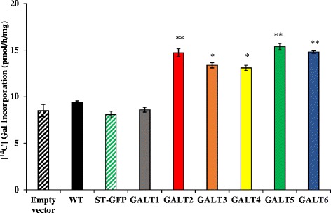
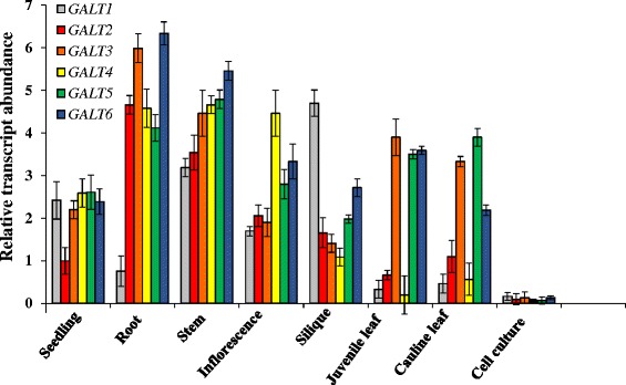
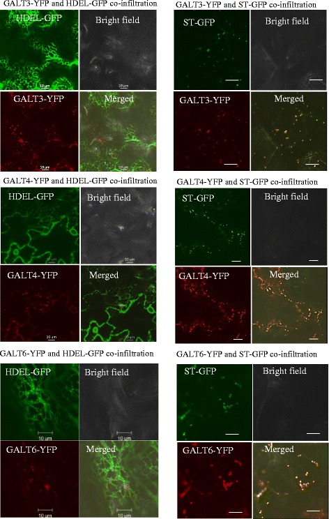
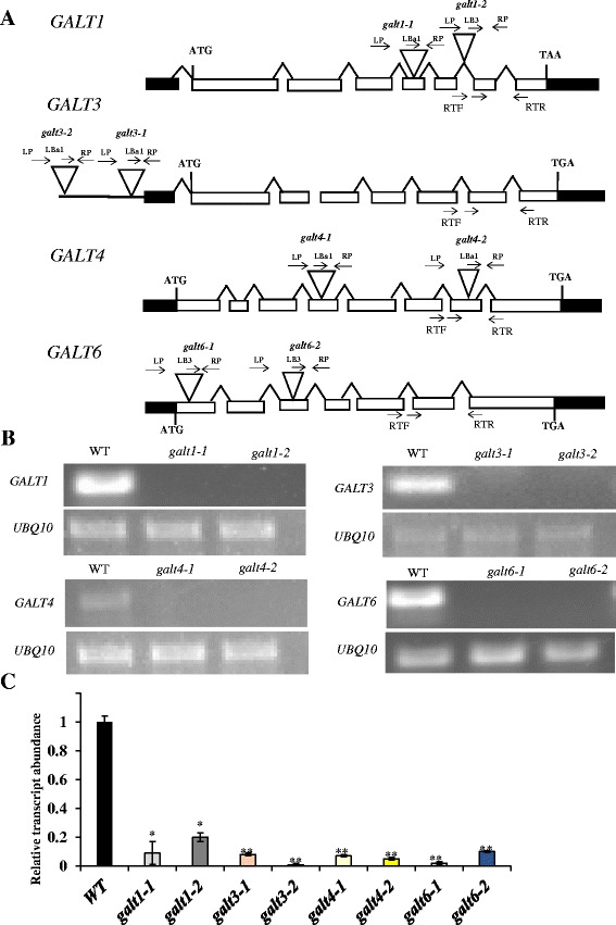
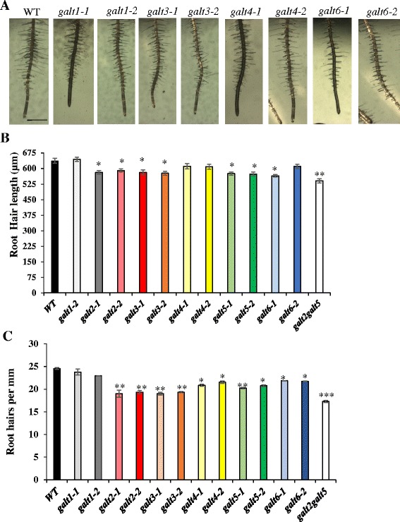
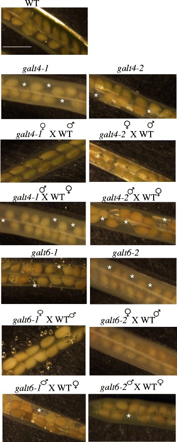
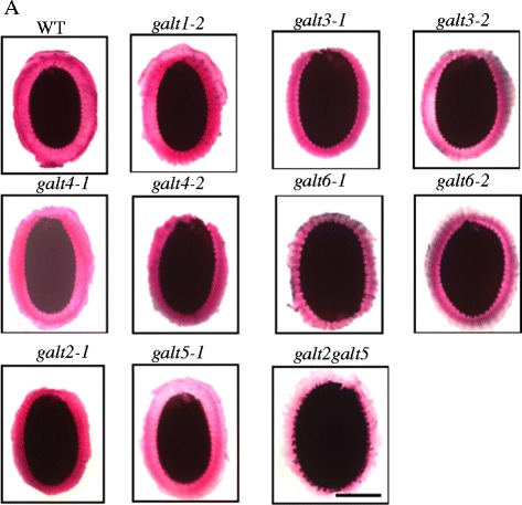
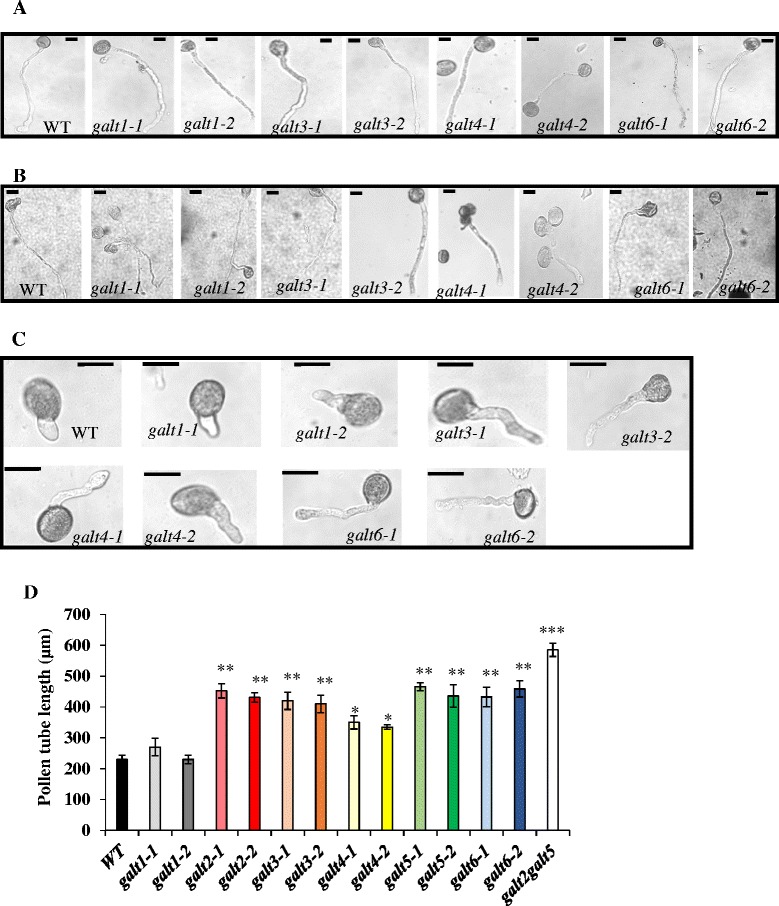
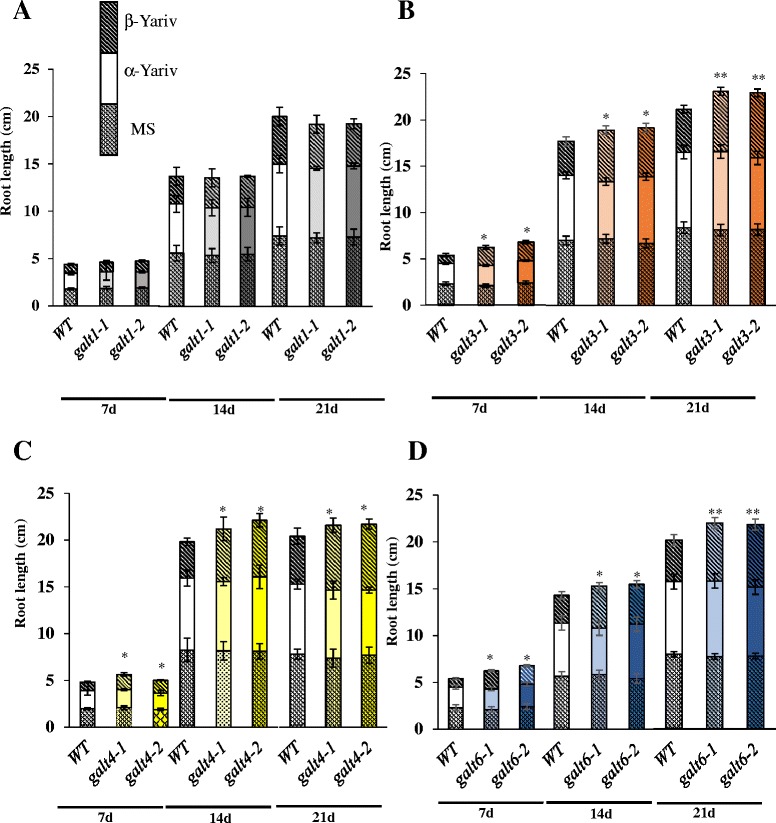
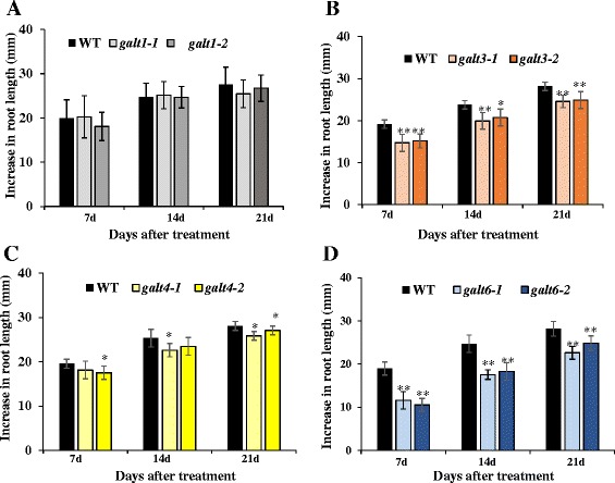
Results: Three members of the GT31 family (GALT3-At3g06440, GALT4-At1g27120, and GALT6-At5g62620) were identified as Hyp-O-GALT genes by heterologous expression in tobacco leaf epidermal cells and examined along with two previously characterized Hyp-O-GALT genes, GALT2 and GALT5. Transcript profiling by real-time PCR of these five Hyp-O-GALTs revealed overlapping but distinct expression patterns. Transiently expressed GALT3, GALT4 and GALT6 fluorescent protein fusions were localized within Golgi vesicles. Biochemical analysis of knock-out mutants for the five Hyp-O-GALT genes revealed significant reductions in both AGP-specific Hyp-O-GALT activity and β-Gal-Yariv precipitable AGPs. Further phenotypic analysis of these mutants demonstrated reduced root hair growth, reduced seed coat mucilage, reduced seed set, and accelerated leaf senescence. The mutants also displayed several conditional phenotypes, including impaired root growth, and defective anisotropic growth of root tips under salt stress, as well as less sensitivity to the growth inhibitory effects of β-Gal-Yariv reagent in roots and pollen tubes.

Conclusions: This study provides evidence that all five Hyp-O-GALT genes encode enzymes that catalyze the initial steps of AGP galactosylation and that AGP glycans play essential roles in both vegetative and reproductive plant growth.


Figures

Fig. 1
Fig. 1
Hyp-O-GALT activity of GALT1-GALT6 transiently expressed in N. tabacum. GALT1-GALT6 were expressed in epidermal cells of tobacco leaves by Agrobacterium-mediated transient expression, which were used for the preparation of Golgi-enriched microsomal membrane proteins for the Hyp-GALT assays. Synthetic peptide [AO]7 was used as substrate acceptor. WT tobacco leaves infiltrated with Agrobacterium GV3101 strain (Empty vector), WT tobacco leaves, and WT tobacco leaves infiltrated with ST fused with GFP were used as controls. Experiments were performed using duplicate samples and data represent the mean ± SD from two independent experiments. Asterisks indicate mean values significantly different from the WT (Dunnett’s test, *P <0.05; **P <0.01)
Fig. 2
Fig. 2
Substrate specificity of transiently expressed GALT2-6. Detergent permeablized tobacco microsomal membranes obtained from transiently expressed GALT2-6 served as the enzyme source in the GALT reactions. Various peptide and polysaccharide acceptor substrates were tested including: 1) [AO]7, [AO]14, and d[AO]51 which contain 7, 14, and 51 [AO] units, respectively, 2) a chemically synthesized extensin peptide (ExtP) containing repetitive SO4 units, 3) [AP]7 which contains 7 [AP] units, 4) Rhamnogalactan I (RGI) from potato pectin, and 5) RG from soybean pectin. Microsomes obtained from WT tobacco leaves infiltrated with empty pMDC32 vector were used as a negative control and depicted as WT. Enzyme reactions were done in triplicate and mean values ± SE are presented. Asterisks indicate values significantly different from the WT (Dunnett’s test, *P <0.05; **P <0.01)
Fig. 3
Fig. 3
Expression patterns of the six membered GALT gene family. qPCR analysis of GALT1-GALT6 expression Arabidopsis organs and cell cultures. Roots were obtained from 14 day old seedlings grown on MS plates with 1 % sucrose and a week old cell suspension culture was used for RNA extraction. The level of expression was calculated relative to the UBQ10 gene (mean ± SE of three biological replicates)
Fig. 4
Fig. 4
Subcellular localization of transiently expressed GALT3-YFP, GALT4-YFP, and GALT6-YFP in N. tabacum. GALT3-YFP, GALT4-YFP, and GALT6-YFP fusion constructions were expressed under the control of the CaMV 35S promoter in N. tabacum. Transiently expressed GALT3-YFP, GALT4-YFP, and GALT6-YFP co-localized with sialyl transferase (ST)-GFP fusion protein (a Golgi marker), but not with HDEL-GFP fusion protein (an ER marker). These constructs were examined by laser-scanning confocal microscopy under fluorescent and white light, and the fluorescent images were merged to observe co-localization. Size bar = 10 μm
Fig. 5
Fig. 5
Schematic gene models, locations of T-DNA mutant insertions, and transcript analysis of GALT1, GALT3, GALT4, and GALT6. a GALT1, GALT3. GALT4 and GALT6 gene structures and T-DNA insertion sites in galt1-1, galt1-2, galt3-1, galt3-2, galt4-1, galt4-2, galt6-1, and galt6-2 mutants. The intron-exon structures of GALT1, GALT3, GALT4, and GALT6 are indicated (introns are drawn as lines and exons as rectangles, with white rectangles representing coding sequences and black rectangles representing UTRs). Sites of T-DNA insertions are marked (triangles) as are the locations of primer sequences (arrows above the genes) used for PCR screening. b RT-PCR analysis of transcripts from rosette leaves of 14-day-old WT (Col-0) and allelic homozygous galt1, galt3, galt4 and galt6 mutant lines. Arrows below the genes in (a) indicate the position of primers (denoted as RTF and RTR) used for RT-PCR analysis of transcript levels. UBQ10 primers were used as internal controls. c Quantitative real-time reverse transcription -PCR (qRT-PCR) analysis was performed to quantify and compare transcript levels of the indicated genes with that of corresponding WT gene. In other words, the relative expression level of the GALT genes in the mutants was compared to WT values, which were set to a value of 1.0 for each of the GALT genes. Asterisks indicate values significantly different from the WT expression of the indicated genes (Dunnett’s test, *P <0.01; **P <0.001)
Fig. 6
Fig. 6
Root hair length and density reduced in the galt3, galt4, and galt6 mutants. a WT, galt1, galt3, galt4, and galt6 plants were grown on MS agar plates for 10 days. Bar = 1 mm. b Quantification of root hair length and c root hair density of the galt mutants. Asterisks indicate significantly reduced root hair length and density compared with WT controls according to Dunnett’s test (*P <0.05; **P <0.01; n >300)
Fig. 7
Fig. 7
Silique morphology of galt4 and galt6 mutant plants along with reciprocal crosses of these mutants to WT plants. Siliques were treated with ethanol to allow for easy observation of the seeds. Absence of ovules is indicated with an asterisk. Bar = 100 μm
Fig. 8
Fig. 8
Pectin staining of seed coat mucilage in wild type, galt1-galt6 single mutants, and galt2galt5 double mutants. Seeds of the indicated genotypes were prehydrated with water for 90 min and stained with ruthenium red to visualize pectin using a Nikon Phot-lab2 microscope coupled with a SPOT RT color CCD camera and SPOT 4.2 analysis software. Bar = 100 μm
Fig. 9
Fig. 9
The galt single mutants demonstrate reduced inhibition of pollen tube growth in the presence of β-Gal-Yariv reagent. a Representative images of pollen tubes from WT, galt1, galt3, galt4, and galt6 mutants after 16 h in pollen germination medium, and b in pollen germination medium supplemented with 30 μM α-Gal-Yariv, and c in pollen germination medium supplemented with 30 μM β-Gal-Yariv reagent. Bar = 50 μm. d Pollen tube lengths from WT, galt1-galt6 mutants, and galt2galt5 double mutants were measured over 16 h in the pollen germination medium supplemented with 30 μM β-Gal-Yariv reagent. Twenty flowers from each genotype and 25 pollen tubes from each flower were measured using Image J software. The experiment was done in triplicate, and the asterisks indicate mean values significantly different from the WT (Dunnett’s test, *P <0.05; **P <0.01; ***P <0.001)
Fig. 10
Fig. 10
Reduced inhibition of primary root growth of galt3, galt4, and galt6 mutants in the presence of β-Gal-Yariv reagent. Root lengths of WT, galt1, galt3, galt4, and galt6 plants were measured 7, 14, and 21 days after germination and seedling establishment for 5 days on MS plates, on MS plates supplemented with 50 μM α-Gal-Yariv reagent, and on MS plates supplemented with 50 μM β-Gal-Yariv reagent. Statistical differences were determined by ANOVA, followed by the Tukey’s honestly significant difference test. Asterisks indicate mean values significantly different from the WT expression of the indicated genes within a treatment group (Dunnett’s test, *P <0.05; **P <0.01). Vertical bars represent mean ± SE of the experimental means from at least three independent experiments, where experimental means were obtained from 10 to 15 seedlings per experiment
Fig. 11
Fig. 11
Salt-induced inhibition of primary root elongation in galt3, galt4, and galt6 mutants. Five-day-old WT, galt1, galt3, galt4, and galt6 seedlings germinated on MS medium were transferred onto media containing 100 mM NaCl and grown vertically. Root elongation (i.e., increase in length after transfer) was measured after 7, 14, and 21 days of growth in the non-permissive media. Data are the means ± SE of measurements from five independent experiments (total n = 100). Asterisks indicate mean values significantly different from the WT (Dunnett’s test, *P <0.05; **P <0.01)

Similar articles

Cited by

References

    1. Majewska-Sawka A, Nothnagel EA. The multiple roles of arabinogalactan proteins in plant development. Plant Physiol. 2000;122:3–10. doi: 10.1104/pp.122.1.3. - DOI - PMC - PubMed
    1. Showalter AM, Keppler B, Lichtenberg J, Gu D, Welch LR. A bioinformatics approach to the identification, classification, and analysis of hydroxyproline-rich glycoproteins. Plant Physiol. 2010;153:485–513. doi: 10.1104/pp.110.156554. - DOI - PMC - PubMed
    1. Ma H, Zhao J. Genome-wide identification, classification, and expression analysis of the arabinogalactan protein gene family in rice (Oryza sativa L.) J Exp Bot. 2010;61:2647–2668. doi: 10.1093/jxb/erq104. - DOI - PMC - PubMed
    1. Seifert GJ, Roberts K. The biology of arabinogalactan proteins. Annu Rev Plant Biol. 2007;58:137–161. doi: 10.1146/annurev.arplant.58.032806.103801. - DOI - PubMed
    1. Ellis M, Egelund J, Schultz CJ, Bacic A. Arabinogalactan proteins: key regulators at the cell surface? Plant Physiol. 2010;153:403–419. doi: 10.1104/pp.110.156000. - DOI - PMC - PubMed

Publication types

MeSH terms