

Longitudinal Diffusion Tensor Imaging Detects Recovery of Fractional Anisotropy Within Traumatic Axonal Injury Lesions
- PMID: 26690938
- PMCID: PMC4884487
- DOI: 10.1007/s12028-015-0216-8
Longitudinal Diffusion Tensor Imaging Detects Recovery of Fractional Anisotropy Within Traumatic Axonal Injury Lesions
Abstract
Background: Traumatic axonal injury (TAI) may be reversible, yet there are currently no clinical imaging tools to detect axonal recovery in patients with traumatic brain injury (TBI). We used diffusion tensor imaging (DTI) to characterize serial changes in fractional anisotropy (FA) within TAI lesions of the corpus callosum (CC). We hypothesized that recovery of FA within a TAI lesion correlates with better functional outcome.
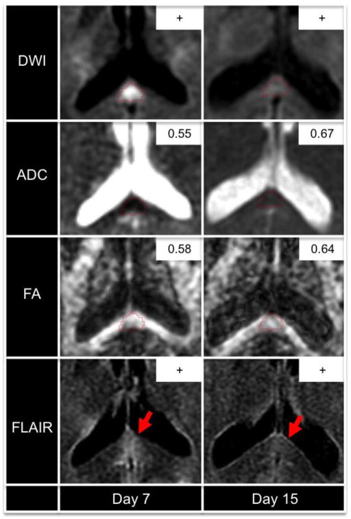
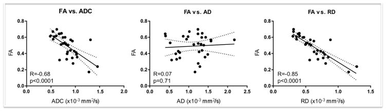
Methods: Patients who underwent both an acute DTI scan (≤day 7) and a subacute DTI scan (day 14 to inpatient rehabilitation discharge) at a single institution were retrospectively analyzed. TAI lesions were manually traced on the acute diffusion-weighted images. Fractional anisotropy (FA), apparent diffusion coefficient (ADC), axial diffusivity (AD), and radial diffusivity (RD) were measured within the TAI lesions at each time point. FA recovery was defined by a longitudinal increase in CC FA that exceeded the coefficient of variation for FA based on values from healthy controls. Acute FA, ADC, AD, and RD were compared in lesions with and without FA recovery, and correlations were tested between lesional FA recovery and functional recovery, as determined by disability rating scale score at discharge from inpatient rehabilitation.
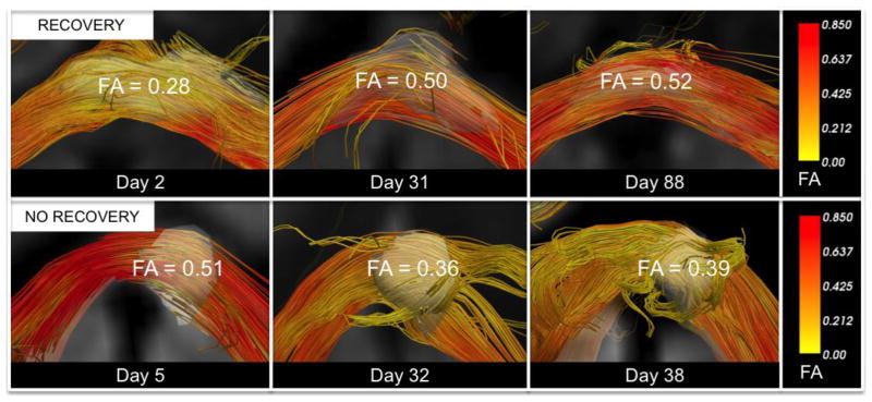
Results: Eleven TAI lesions were identified in 7 patients. DTI detected FA recovery within 2 of 11 TAI lesions. Acute FA, ADC, AD, and RD did not differ between lesions with and without FA recovery. Lesional FA recovery did not correlate with disability rating scale scores.
Conclusions: In this retrospective longitudinal study, we provide initial evidence that FA can recover within TAI lesions. However, FA recovery did not correlate with improved functional outcomes. Prospective histopathological and clinical studies are needed to further elucidate whether lesional FA recovery indicates axonal healing and has prognostic significance.
Keywords: Apparent diffusion coefficient; Diffusion tensor imaging; Fractional anisotropy; Traumatic axonal injury; Traumatic brain injury.
Figures
References
-
- Gentry LR, Godersky JC, Thompson B. MR imaging of head trauma: review of the distribution and radiopathologic features of traumatic lesions. AJR Am J Roentgenol. 1988;150:663–72. - PubMed
-
- Adams JH, Jennett B, Murray LS, et al. Neuropathological findings in disabled survivors of a head injury. J Neurotrauma. 2011;28:701–9. - PubMed
-
- Blumbergs PC, Scott G, Manavis J, et al. Staining of amyloid precursor protein to study axonal damage in mild head injury. Lancet. 1994;344:1055–6. - PubMed
-
- Smith DH, Meaney DF, Shull WH. Diffuse axonal injury in head trauma. J Head Trauma Rehabil. 2003;18:307–16. - PubMed
-
- Christman CW, Grady MS, Walker SA, et al. Ultrastructural studies of diffuse axonal injury in humans. J Neurotrauma. 1994;11:173–86. - PubMed
Publication types
MeSH terms
Grants and funding
LinkOut - more resources
Full Text Sources
Other Literature Sources
Medical
Miscellaneous
