. 2015 Dec 22:5:17559.
doi: 10.1038/srep17559.

Identification of PCSK9 as a novel serum biomarker for the prenatal diagnosis of neural tube defects using iTRAQ quantitative proteomics

Affiliations

Identification of PCSK9 as a novel serum biomarker for the prenatal diagnosis of neural tube defects using iTRAQ quantitative proteomics

Dong An et al. Sci Rep. .

Abstract

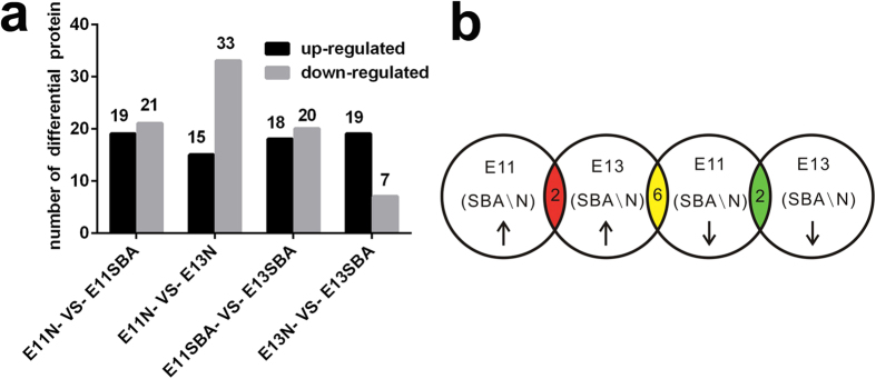
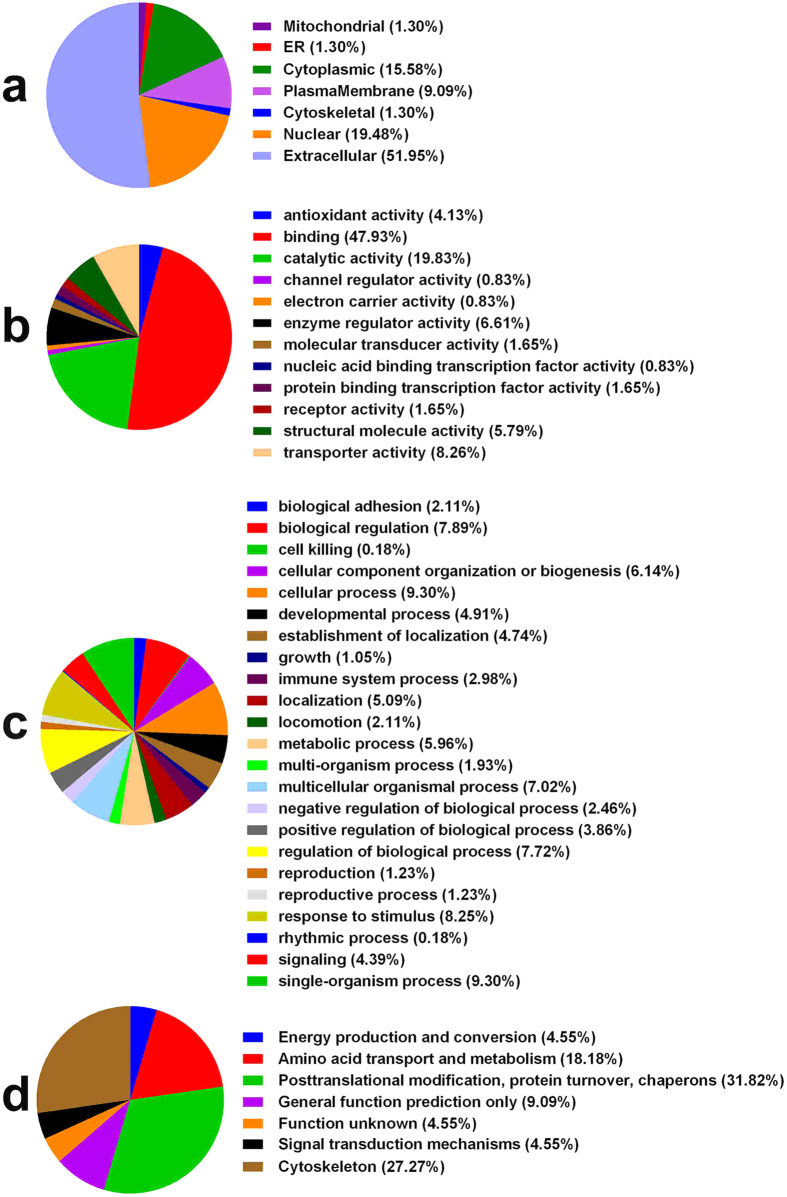
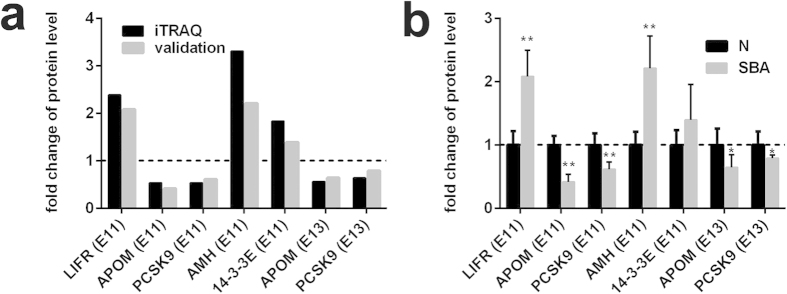
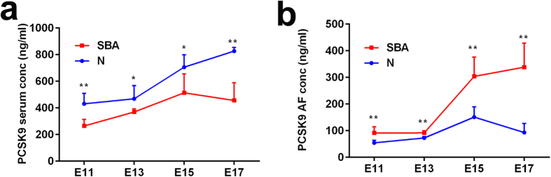
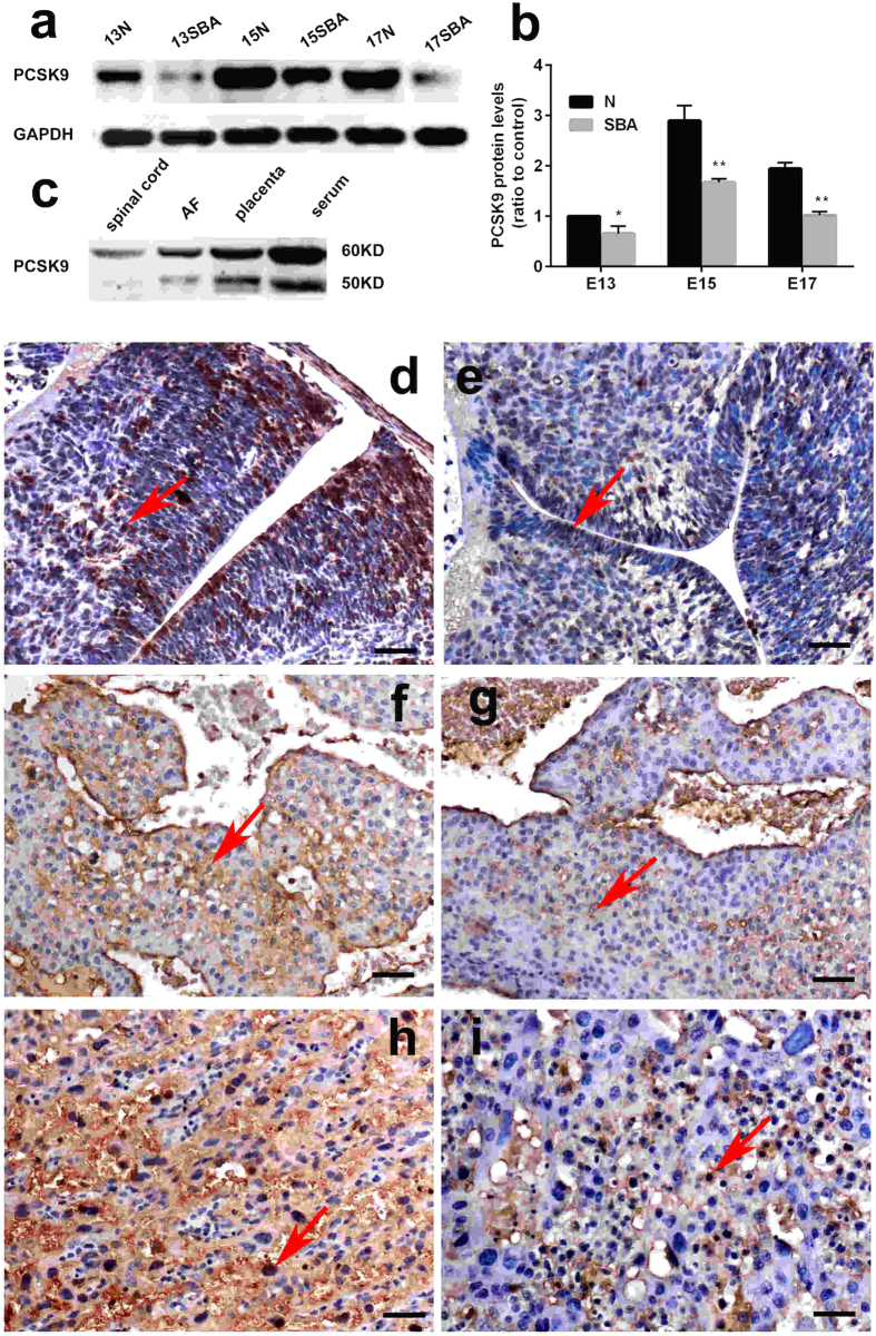
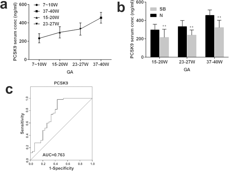
To identify candidate serum molecule biomarkers for the non-invasive early prenatal diagnosis of neural tube defects (NTDs), we employed an iTRAQ-based quantitative proteomic approach to analyze the proteomic changes in serum samples from embryonic day (E) 11 and E13 pregnant rats with spina bifida aperta (SBA) induced by all-trans retinoic acid. Among the 390 proteins identified, 40 proteins at E11 and 26 proteins at E13 displayed significant differential expression in the SBA groups. We confirmed 5 candidate proteins by ELISA. We observed the space-time expression changes of proprotein convertase subtilisin/kexin type 9 (PCSK9) at different stages of fetal development, including a marked decrease in the sera of NTD pregnancies and gradual increase in the sera of normal pregnancies with embryonic development. PCSK9 demonstrated the diagnostic efficacy of potential NTD biomarkers [with an area under the receiver operating characteristic curve of 0.763, 95% CI: 065-0.88]. Additionally, PCSK9 expression in the spinal cords and placentas of SBA rat fetuses was markedly decreased. PCSK9 could serve as a novel molecular biomarker for the non-invasive prenatal screening of NTDs and may be involved in the pathogenesis of NTDs at critical periods of fetal development.


Figures

Figure 1
Figure 1. Differentially expressed serum proteins between pregnant rats with SBA fetuses and normal controls at E11 and E13.
(a) The number of differentially expressed serum proteins between pregnant rats with SBA fetuses and normal controls at E11 and E13. (b) Distribution of the up-regulated and down-regulated expression of differential proteins at E11 and E13; the yellow region includes 6 proteins (sPLA2, fibroleukin, GDN, tubulin beta chain, fibrinogen-like protein 1, haptoglobin) that were up-regulated at E13 and down-regulated at E11, the green region includes 2 proteins (APOM, PCSK9) that were down-regulated at both E11 and E13, and the red region includes 2 proteins (fibrinogen gamma chain, Ig heavy chain V region MOPC 47A) that were up-regulated at both E11 and E13.
Figure 2
Figure 2. GO annotation and functional classification of differentially expressed serum proteins: Gene ontology terms for subcellular location distribution (a), molecular functions (b), biological process (c) and COG Function Classification (d).
Figure 3
Figure 3. Expression patterns of selected protein candidates in the serum of the experimental group compared with the control group using iTRAQ analysis and ELISA validation.
(a) Fold change of protein levels (the mean value of experimental group / the mean value of control group) of LIFR (E11), AMH (E11), 14-3-3E (E11), APOM (E11 and E13), and PCSK9 (E11 and E13) from iTRAQ analysis and ELISA validation. (b) Fold change of protein levels (the protein concentration of each sample/the mean concentration of control group) of LIFR (E11), AMH (E11), 14-3-3E (E11), APOM (E11 and E13), and PCSK9 (E11 and E13) from ELISA analysis (n = 6). *p < 0.05, **p < 0.01 (pregnant rats with SBA serum vs. control serum). The data are shown as the mean value  ±  SD. The upregulated proteins in the experimental group were above the dotted line, and the downregulated were below the dotted line.
Figure 4
Figure 4. Serum and AF levels of PCSK9 in pregnant rat with SBA fetuses and normal fetuses for different time points.
(a) Serum levels of PCSK9 in pregnant rat with SBA fetuses and normal fetuses for different time points. (b) AF Levels of PCSK9 in pregnant rats with SBA fetuses and normal fetuses for different time points. Six rats were used as a group from E11 to E17 (n = 6). *p < 0.05, **p < 0.01 (pregnant rats with SBA fetuses vs. control group). The data are shown as the mean value  ±  SD.
Figure 5
Figure 5. Western blot and IHC analysis of PCSK9 in the spinal cords and placentas of rat embryos.
(a,b) Western blot analysis showed decreased levels of PCSK9 in the SBA rat fetuses (p < 0.001). (c) PCSK9 was detectable in serum, AF and the spinal cord and placenta by Western blot analysis. Protein extracts revealed one band (60 kDa) in spinal cord and two bands (60, 50 kDa) in serum, AF and placenta. (d–i) Distribution of PCSK9 immunostaining in the spinal cords and placentas of rat embryos at E15. PCSK9 immunoreactivity was mainly localized to the cytoplasm and nuclear (red arrow) in the spinal cords and the placentas. PCSK9 showed downregulated expression in SBA embryos compared with normal ones. (d) Spinal cord of normal embryos. (e) Spinal cord of SBA embryos. (f) The maternal side of placenta of normal embryos. (g) The maternal side of placenta of SBA embryos. (h) The fetal side of a placenta of normal embryos. (i) The fetal side of a placenta of SBA embryos. Magnification: ×200; Scale bars = 50 μm.
Figure 6
Figure 6. Levels of PCSK9 in NTD maternal serum.
(a) PCSK9 concentrations in the serum of women with healthy fetuses at different GAs. (b) PCSK9 levels in the sera of women with fetuses with NTDs and healthy fetuses at different GAs (GA 15–20 w, 23–27 w, 37–40 w). (c) ROC analysis for PCSK9 to discriminate NTDs from controls. **p < 0.01.

References

    1. Molloy A. M. & Kappen C. Papers from the 7th International Neural Tube Defects Conference. Birth Defects Res A Clin Mol Teratol 94, 747–748 (2012). - PubMed
    1. Copp A. J., Greene N. D. & Murdoch J. N. The genetic basis of mammalian neurulation. Nat Rev Genet 4, 784–793 (2003). - PubMed
    1. Brock D. J. & Scrimgeour J. B. Early prenatal diagnosis of anencephaly. Lancet 2, 1252–1253 (1972). - PubMed
    1. Seller M. J., Campbell S., Coltart T. M. & Singer J. D. Early termination of anencephalic pregnancy after detection by raised alpha-fetoprotein levels. Lancet 2, 73 (1973). - PubMed
    1. Flick A., Krakow D., Martirosian A., Silverman N. & Platt L. D. Routine measurement of amniotic fluid alpha-fetoprotein and acetylcholinesterase: the need for a reevaluation. Am J Obstet Gynecol 211, 139 e131–136 (2014). - PubMed

Publication types

MeSH terms