. 2016 Apr;17(2):81-8.
doi: 10.1007/s10162-015-0551-7. Epub 2015 Dec 21.

Increased Spontaneous Otoacoustic Emissions in Mice with a Detached Tectorial Membrane

Affiliations

Increased Spontaneous Otoacoustic Emissions in Mice with a Detached Tectorial Membrane

Mary Ann Cheatham et al. J Assoc Res Otolaryngol. 2016 Apr.

Abstract

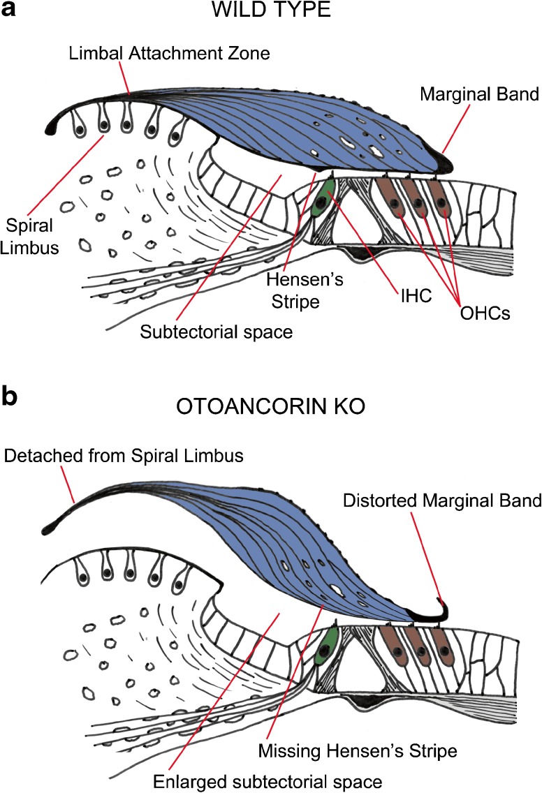
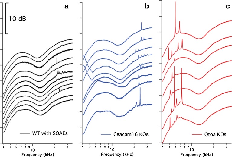
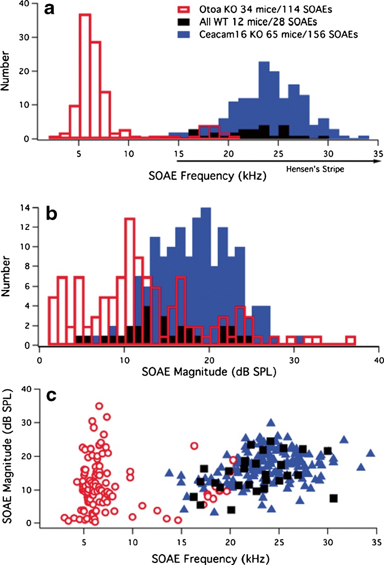
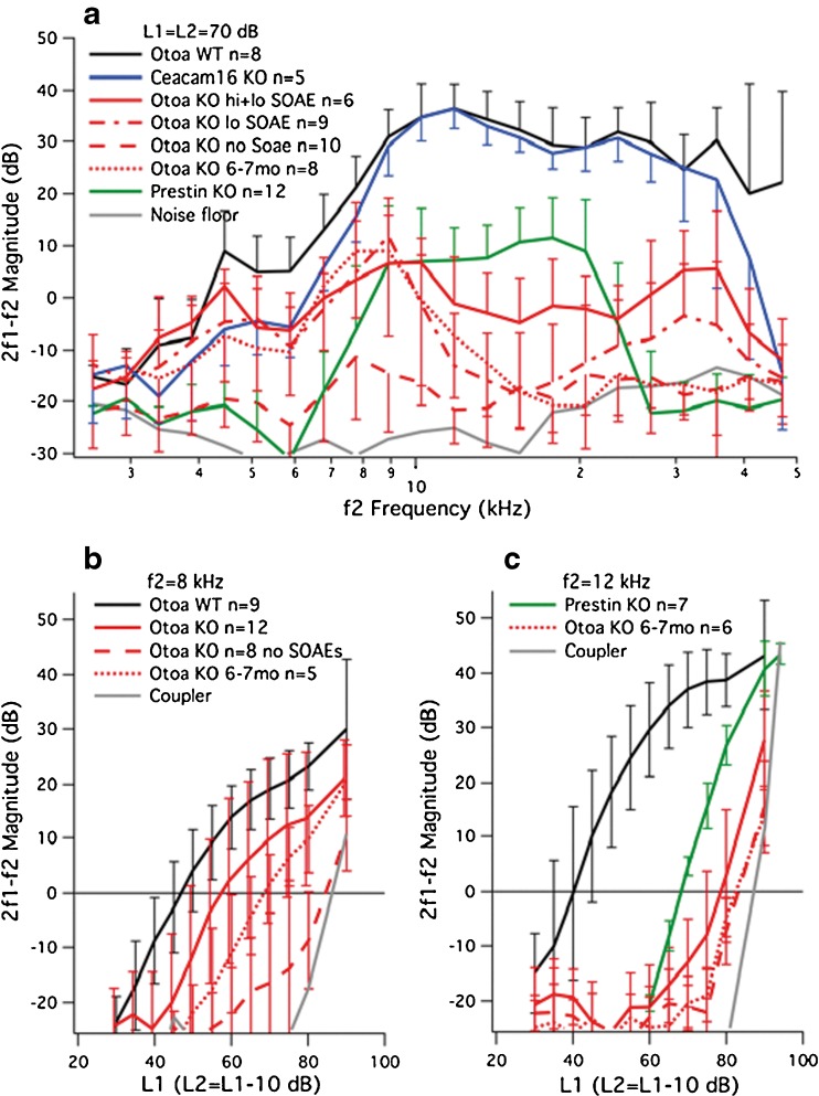
Mutations in genes encoding tectorial membrane (TM) proteins are a significant cause of human hereditary hearing loss (Hildebrand et al. 2011), and several mouse models have been developed to study the functional significance of this accessory structure in the mammalian cochlea. In this study, we use otoacoustic emissions (OAE), signals obtained from the ear canal that provide a measure of cochlear function, to characterize a mouse in which the TM is detached from the spiral limbus due to an absence of otoancorin (Otoa, Lukashkin et al. 2012). Our results demonstrate that spontaneous emissions (SOAE), sounds produced in the cochlea without stimulation, increase dramatically in mice with detached TMs even though their hearing sensitivity is reduced. This behavior is unusual because wild-type (WT) controls are rarely spontaneous emitters. SOAEs in mice lacking Otoa predominate around 7 kHz, which is much lower than in either WT animals when they generate SOAEs or in mutant mice in which the TM protein Ceacam16 is absent (Cheatham et al. 2014). Although both mutants lack Hensen's stripe, loss of this TM feature is only observed in regions coding frequencies greater than ~15 kHz in WT mice so its loss cannot explain the low-frequency, de novo SOAEs observed in mice lacking Otoa. The fact that ~80 % of mice lacking Otoa produce SOAEs even when they generate smaller distortion product OAEs suggests that the active process is still functioning in these mutants but the system(s) involved have become less stable due to alterations in TM structure.

Keywords: Hensen’s stripe; active process; cochlea; otoancorin; spontaneous otoacoustic emissions; tectorial membrane.


Figures

Fig. 1
Fig. 1
Tectorial membrane anatomy. Diagram showing normal (A) and detached (B) tectorial membranes in WT controls and Otoa KO mice. The drawings of the organ of Corti are based on anatomical results published by Lukashkin et al. (2012).
Fig. 2
Fig. 2
SOAE spectra. Representative SOAE spectra are provided for all WT animals (A) in our collection, along with those from a selection of mice lacking Ceacam16 (B) or otoancorin (C). All data are plotted on an ordinate with arbitrary scale, i.e., the 10-dB scale applies to all panels.
Fig. 3
Fig. 3
SOAE frequencies and magnitudes. The number of SOAEs is plotted as a function of their peak frequency (A) and magnitude (B). The total number of mice with SOAEs and the total number of SOAEs observed in each mouse group is provided in the legend. The position of Hensen’s stripe in WT mice is also indicated in panel A along the abscissa to show that it is present in controls but only for more basal locations encoding frequencies greater than ~15 kHz (Cheatham et al. ; Legan et al. 2014). Panel C provides a plot of SOAE magnitude versus SOAE frequency for WT (black squares), Ceacam16 KOs (blue triangles), and Otoa KOs (red open circles).
Fig. 4
Fig. 4
DPOAEs. Panel A provides DPOAE magnitudes for 2f1-f2 plotted as a function of f2 frequency for equal-level primaries at 70 dB. Although Ceacam16 KO mice (blue) have near-normal responses when young, the Otoa KOs show reduced responses. The animals lacking Otoa are plotted in red for four different groups: mice producing both high- (hi) and low- (lo) frequency SOAEs with solid lines, those generating low-frequency SOAEs only with dot-dashed lines, those lacking SOAEs with dashed lines, and Otoa KO mice at 6–7 months of age with dotted lines. Results from mice lacking prestin are also appended and plotted in green. Data are presented as means and standard deviations (plotted in one direction only for clarity), and the controls are plotted in black. It is also emphasized that in our hands the Otoa KO mice show smaller DPOAEs than in the initial report on this mouse mutant (Lukashkin et al. 2012). This difference may reflect strain background such that the data reported here are for mice on a mixed, variable 129/B6 background, while those used by Lukashkin et al. were CBA/Ca × B6 F1 hybrids. The backcross to CBA/CaJ appears to minimize the age-related hearing loss known to plague the C57BL6J line. Input-output functions for 2f1-f2 are plotted in panel B for f2 = 8 kHz and in panel C for f2 = 12 kHz. Data for Otoa KO mice in panels B and C are again separated into two groups: those with (red solid lines) and those without (red dashed lines) SOAEs. Input-output functions for older Otoa KO mice are also appended and appear as dotted lines. Panel C also shows results from prestin KO mice (green). Distortion in the sound was measured in a tubing coupler and is indicated in panels B and C by the solid gray line.

References

    1. Carvalho S, Mom T, Gilain L, Avan P. Frequency specificity of distortion-product otoacoustic emissions produced by high-level tones despite inefficient cochlear electromechanical feedback. J Acous Soc Am. 2004;116:1639. doi: 10.1121/1.1777873. - DOI - PubMed
    1. Cheatham MA, Goodyear RJ, Homma K, Legan PK, Korchagina J, Naskar S, Siegel JH, Dallos P, Zheng J, Richardson GP. Loss of the tectorial membrane protein CEACAM16 enhances spontaneous, stimulus-frequency, and transiently evoked otoacoustic emissions. J Neurosci. 2014;34:10325–10338. doi: 10.1523/JNEUROSCI.1256-14.2014. - DOI - PMC - PubMed
    1. Crawford AC, Fettiplace R. The mechanical properties of ciliary bundles of turtle cochlear hair cells. J Physiol. 1985;364:359–379. doi: 10.1113/jphysiol.1985.sp015750. - DOI - PMC - PubMed
    1. Dallos P, Billone MC, Durrant JD, Wang C, Raynor S. Cochlear inner and outer hair cells: functional differences. Science. 1972;177:356–358. doi: 10.1126/science.177.4046.356. - DOI - PubMed
    1. Dallos P, Wu X, Cheatham MA, Gao J, Zheng J, Anderson CT, Jia S, Wang X, Cheng WH, Sengupta S, He DZ, Zuo J. Prestin-based outer hair cell motility is necessary for mammalian cochlear amplification. Neuron. 2008;58:1–7. doi: 10.1016/j.neuron.2008.02.028. - DOI - PMC - PubMed

Publication types

LinkOut - more resources