Review
. 2015 Nov-Dec;20(6):110-25.
doi: 10.1590/2177-6709.20.6.110-125.sar.

Facial asymmetry: a current review

Affiliations
Review

Facial asymmetry: a current review

Guilherme Thiesen et al. Dental Press J Orthod. 2015 Nov-Dec.

Abstract

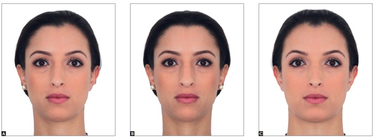
The term "asymmetry" is used to make reference to dissimilarity between homologous elements, altering the balance between structures. Facial asymmetry is common in the overall population and is often presented subclinically. Nevertheless, on occasion, significant facial asymmetry results not only in functional, but also esthetic issues. Under these conditions, its etiology should be carefully investigated in order to achieve an adequate treatment plan. Facial asymmetry assessment comprises patient's first interview, extra- as well as intraoral clinical examination, and supplementary imaging examination. Subsequent asymmetry treatment depends on patient's age, the etiology of the condition and on the degree of disharmony, and might include from asymmetrical orthodontic mechanics to orthognathic surgery. Thus, the present study aims at addressing important aspects to be considered by the orthodontist reaching an accurate diagnosis and treatment plan of facial asymmetry, in addition to reporting treatment of some patients carriers of such challenging disharmony.

O termo assimetria é utilizado quando existe uma desigualdade entre as partes homólogas, afetando, assim, o equilíbrio entre as estruturas. A assimetria facial é comum na população, e muitas vezes se apresenta de forma subclínica. Entretanto, em alguns casos existe uma assimetria facial significativa que resulta tanto em problemas funcionais quanto estéticos. Nessas circunstâncias, a etiologia deve ser cuidadosamente investigada, para que seja possível elaborar um plano de tratamento adequado. A avaliação da assimetria facial é constituída por uma anamnese do paciente, exame clínico extra e intrabucal, além de exames complementares de imagem. O tratamento subsequente dessa assimetria depende da idade do paciente, etiologia e da magnitude da desarmonia, podendo envolver desde mecânicas ortodônticas assimétricas até a realização de cirurgia ortognática. Assim, o presente artigo busca abordar aspectos importantes para que o ortodontista possa realizar um adequado diagnóstico e plano de tratamento da assimetria facial, além de relatar o tratamento de alguns pacientes portadores dessa desafiadora desarmonia.


Figures

Figure 1
Figure 1. - Extraoral photograph in frontal view. Patient with relative symmetry (A) in which the median sagittal plane was determined and used as the basis to create mirror images on the right and left sides (B and C, respectively). Note that the original and mirror images differ from one another.
Figure 2
Figure 2. - Class II growing patient with mandibular deficiency. Presence of mild facial asymmetry with deviation of the chin to the left. Initial extraoral (A, B and C) and intraoral photographs (D, E, F, G and H), as well as profile, panoramic and carpal radiographs (I, J and K).
Figure 3
Figure 3. - Telescopic mechanism of the Herbst appliance in place. Asymmetrical mandibular advancement aiming at correcting skeletal occlusal and facial asymmetry. Lateral intraoral photographs on the right side (A), in frontal view (B) and on the left side (C).
Figure 4
Figure 4. - Treatment outcomes for the patient presented in Figure 2, after the second phase of treatment conducted with full fixed orthodontic appliance. Final extraoral (A, B and C) and intraoral (D, E, F, G and H) photographs. Profile and panoramic radiographs (I and J).
Figure 5
Figure 5. - Patient presenting Class II malocclusion on the right side, with negative cephalometric discrepancy and discreet crowding in both arches. Mild facial asymmetry with deviation of the mandible to the right. Initial extraoral (A, B and C) and intraoral photographs (D, E, F, G and H), as well as profile and panoramic radiographs (I and J).
Figure 6
Figure 6. - Corrective orthodontic treatment with protocol including three extractions (teeth #14,24 and 34). Extraction in the mandibular arch was recommended for correction of lower dental midline coinciding with patient's median sagittal plane, in addition to correcting protrusion and crowding of mandibular anterior teeth. Extractions in the maxillary arch were carried out to correct protrusion, crowding and overjet. Lateral intraoral photographs on the right side (A), in frontal view (B) and on the left side (C).
Figure 7
Figure 7. - Treatment outcomes for the patient presented in Figure 5. Final extraoral (A, B and C) and intraoral (D, E, F, G and H) photographs. Profile and panoramic radiographs (I and J).
Figure 8
Figure 8. - Class I mature patient with asymmetry evinced by lateral deviation of the chin, in addition to vertical difference in leveling between lip commissures and inclination of the occlusal plane in frontal view. Initial extraoral (A, B and C) and intraoral photographs (D, E, F, G and H), as well as profile, posterior-anterior and panoramic radiographs (I, J and K).
Figure 9
Figure 9. - Clinical aspect after presurgical orthodontic preparation carried out with a view to correcting dental tipping at their basal bones. The three planes of space must be considered. Extraoral (A, B, C and D) and intraoral photographs (E, F and G), as well as CT scans with soft tissues overlapping hard tissues (H, I and J).
Figure 10
Figure 10. - Treatment outcomes for the patient presented in Figure 8. Final extraoral (A, B and C) and intraoral (D, E, F, G, H and I) photographs. Profile, posterior-anterior and panoramic radiographs (J, K and L).
Figure 11
Figure 11. - Tomographic superimposition of patient presented in Figures 8 to 10 evincing changes before and after surgical correction of facial asymmetry (A, B and C). Surgical maxillary advancement of 4 mm was carried out, in addition to 1.5-mm impaction in the anterior region, 2-mm asymmetrical impaction in the posterior region on the right side and 2.5-mm asymmetrical impaction in the posterior region on the left side. The mandible was rotated for asymmetry correction

References

    1. Lindauer SJ. Asymmetries: diagnosis and treatment (editorial) Semin Orthod. 1998;4(3):133–133. - PubMed
    1. Cheong YW, Lo LJ. Facial asymmetry: etiology, evaluation, and management. Chang Gung Med J. 2011 Jul-Aug;34(4):341–351. - PubMed
    1. IPDTOC Working Group Prevalence at birth of cleft lip with or without cleft palate: data from the International Perinatal Database of Typical Oral Clefts (IPDTOC) Cleft Palate Craniofac J. 2011;48(1):66–81. - PubMed
    1. Peck S, Peck L, Kataja M. Skeletal asymmetry in esthetically pleasing faces. Angle Orthod. 1991;61(1):43–48. - PubMed
    1. Rhodes G, Sumich A, Byatt G. Are Average facial configurations attractive only because of their symmetry. Psychol Sci. 1999;10(1):52–58.