. 2016 Feb;48(2):152-8.
doi: 10.1038/ng.3475. Epub 2015 Dec 21.

Regulatory mutations in TBX3 disrupt asymmetric hair pigmentation that underlies Dun camouflage color in horses

Affiliations

Regulatory mutations in TBX3 disrupt asymmetric hair pigmentation that underlies Dun camouflage color in horses

Freyja Imsland et al. Nat Genet. 2016 Feb.

Abstract

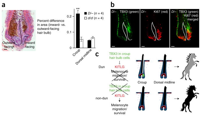
Dun is a wild-type coat color in horses characterized by pigment dilution with a striking pattern of dark areas termed primitive markings. Here we show that pigment dilution in Dun horses is due to radially asymmetric deposition of pigment in the growing hair caused by localized expression of the T-box 3 (TBX3) transcription factor in hair follicles, which in turn determines the distribution of hair follicle melanocytes. Most domestic horses are non-dun, a more intensely pigmented phenotype caused by regulatory mutations impairing TBX3 expression in the hair follicle, resulting in a more circumferential distribution of melanocytes and pigment granules in individual hairs. We identified two different alleles (non-dun1 and non-dun2) causing non-dun color. non-dun2 is a recently derived allele, whereas the Dun and non-dun1 alleles are found in ancient horse DNA, demonstrating that this polymorphism predates horse domestication. These findings uncover a new developmental role for T-box genes and new aspects of hair follicle biology and pigmentation.


Conflict of interest statement

COMPETING FINANCIAL INTERESTS

The authors declare no competing financial interests.

Figures

Figure 1
Figure 1
Phenotypic characterization. (a) Three horses with different genotypes at the Dun g locus on a similar pigmentary (E/−; a/a) background, including a blue Dun (D/−) horse, a black horse with primitive markings (d1/d1) and a black horse without primitive markings (d2/d2). Photographs by Freyja Imsland and Páll Imsland. (b) Cross-sections of hairs from the croup of the three horses in a. (c) Skin and hair section stained with hematoxylin and eosin from a Dun horse. (d) Hair sections stained with hematoxylin and eosin from Dun and non-dun horses. (e) Color intensity differences across the diameter of the hair cortex (means ± s.e.m.) as shown in the inset for the croup and dorsal midline in each phenotype (n = 6 Dun and n = 6 non-dun). **P < 0.01 for Dun versus non-dun croup, two-tailed t test. (f) Cladogram of the Equidae family (based on ref. 10); species with hair histology in g are shown in bold. (g) Photographs of Przewalski’s horses (Equus ferus przewalskii) and Somali wild ass (Equus africanus somaliensis) (left) and photomicrographs of transverse sections through dilute-colored flank hairs (right). Photographs by Waltraut Zimmerman and the St. Louis Zoo. Scale bars, 35 μm (b), 100 μm (c), 35 μm (d), 10 μm (e) and 50 μm (g).
Figure 2
Figure 2
Genetic analysis. (a) Association analysis (χ2) of the Dun phenotype with SNP genotypes at chr. 8: 18,061,745–18,482,196. (b) Read alignments from whole-genome sequencing of a Dun heterozygote and a non-dun homozygote. Red borders denote a read pair where one of the reads is unmapped; blue segments represent soft-clipped parts of reads where part of the read cannot be aligned. The position of the deletion downstream of TBX3 at chr. 8: 18,227,267–18,227,279 is indicated, and the extent of sequence conservation is illustrated using human genome annotation. (c) Alignment of the Dun, non-dun1 and non-dun2 alleles at the deletion breakpoints, showing how the deletion breakpoint closer to TBX3 is flanked by an additional deletion of 8 bp in non-dun2; there is a 1-bp indel polymorphism between Dun and non-dun1. (d) SNP association analysis (−log10 (P value), two-tailed Fisher’s exact test) between Dun and non-dun1 haplotypes, including both domestic and Przewalski’s horses. SNP1 and SNP2 are marked with a black arrow. Red dots mark candidate SNPs selected on the basis of allele frequencies of more than 50% in Dun individuals (dominant trait) and less than 5% in non-dun individuals (erroneous calls) (supplementary table 3). The location of the deletion is marked with a red arrow in d and f. (e) Alignment of six sequence polymorphisms associated with Dun and non-dun1 haplotypes from domestic horses. SNP1 and SNP2 are shown in bold. (f) Nucleotide diversity at polymorphic sites estimated in sliding windows of 100 SNPs for Dun (D), non-dun1 (d1) and non-dun2 (d2) chromosomes among domestic horses and Przewalski’s horses.
Figure 3
Figure 3
Differential gene and protein expression in the croup skin of Dun and non-dun horses. (a) Transcript levels (normalized gene counts) plotted as a function of differential expression (log2-transformed fold change) in Dun (D/−, n = 7) versus non-dun (d1/d1, n = 3; d2/d2, n = 6; d1/d2, n = 2) samples. The 57 genes demonstrating significant (false discovery rate (FDR) < 0.1) differential expression are shown in dark yellow (higher expression in Dun) or red (lower expression in Dun; Online Methods and Supplementary Table 6); seven additional pigmentation-related genes that are not differentially expressed are shown in blue. (be) Immunofluorescence for TBX3 (green) (b), MITF (green) (c), KIT (green) (d) and KITLG (green) (e) in sections of anagen hair follicles from the croup of Dun/−, non-dun1/non-dun1 and non-dun2/non-dun2 horses. Pigment is pseudocolored in b (red). Corresponding bright-field images are on the right in ce. DAPI staining is in blue in ce, and white lines indicate the basement membrane. Each photomicrograph is representative of at least two individuals of each genotype. Scale bars, 100 μm (be).
Figure 4
Figure 4
Relationship between TBX3 expression and hair follicle symmetry and differentiation. (a) Left, hair follicle section stained with hematoxylin and eosin from Dun croup skin. Black lines outline the outward-facing and inward-facing halves of the hair bulb. Right, the percent difference in area (mean ± s.e.m.) between the outward- and inward-facing halves of the hair bulb was calculated for the croup and dorsal midline from Dun (D/−; n = 4) and non-dun (d/d; n = 4) horses. Bulb area was measured from at least two serial sections (range of 2–12 sections/follicle) from at least two follicles (range of 2–7 follicles) per anatomical location. **P = 0.004 for Dun versus non-dun croup, two-tailed t test. (b) Immunofluorescence for TBX3 (green; left) and Ki67 (red; center) in a croup hair follicle from a Dun horse. The merged image is on the right; white lines delineate the basement membrane. Scale bars, 25 μm (a) and 50 μm (b). (c) Asymmetric expression of TBX3 (green) in hair bulb keratinocytes of Dun croup hairs impairs the expression of KITLG (red) and melanocyte survival in one-half of the follicle. Dun croup hairs are asymmetrically pigmented and appear lighter colored than hairs from the dorsal stripe or from non-dun horses.

References

    1. Wade CM, et al. Genome sequence, comparative analysis, and population genetics of the domestic horse. Science. 2009;326:865–867. - PMC - PubMed
    1. Orlando L, et al. Recalibrating Equus evolution using the genome sequence of an early Middle Pleistocene horse. Nature. 2013;499:74–78. - PubMed
    1. Bowling AT, Ryder OA. Genetic studies of blood markers in Przewalski’s horses. J Hered. 1987;78:75–80. - PubMed
    1. Adalsteinsson S. Inheritance of yellow dun and blue dun in the Icelandic toelter horse. J Hered. 1978;69:146–148. - PubMed
    1. Bamshad M, et al. Mutations in human TBX3 alter limb, apocrine and genital development in ulnar-mammary syndrome. Nat Genet. 1997;16:311–315. - PubMed

Publication types

Substances

LinkOut - more resources