. 2016 Apr;46(4):912-8.
doi: 10.1002/eji.201546075. Epub 2016 Jan 18.

Characterization of a conditional interleukin-1 receptor 1 mouse mutant using the Cre/LoxP system

Affiliations

Characterization of a conditional interleukin-1 receptor 1 mouse mutant using the Cre/LoxP system

Wesam H Abdulaal et al. Eur J Immunol. 2016 Apr.

Abstract

IL-1 is a key cytokine known to drive chronic inflammation and to regulate many physiological, immunological, and neuroimmunological responses via actions on diverse cell types of the body. To determine the mechanisms of IL-1 actions as part of the inflammatory response in vivo, we generated a conditional IL-1 receptor 1 (IL-1R1) mouse mutant using the Cre/LoxP system (IL-1R1(fl/fl) ). In the mutant generated, exon 5, which encodes part of the extracellular-binding region of the receptor, is flanked by LoxP sites, thereby inactivating the two previously described functional IL-1R1 gene transcripts after Cre-mediated recombination. Using keratin 14-Cre driver mice, new IL-1R1 deficient (-/-) mice were subsequently generated, in which all signaling IL-1 receptor isoforms are deleted ubiquitously. Furthermore, using vav-iCre driver mice, we deleted IL-1 receptor isoforms in the hematopoietic system. In these mice, we show that both the IL-17 and IL-22 cytokine response is reduced, when mice are challenged by the helminth Trichuris muris. We are currently crossing IL-1R1(fl/fl) mice with different Cre-expressing mice in order to study mechanisms of acute and chronic inflammatory diseases.

Keywords: Cre/loxP; IL-17; Immune regulation; Infection; Trichuris muris.


Figures

Figure 1
Figure 1
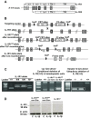
Generation of IL‐1R1fl/fl mice and conditional deletion of IL‐1R1. (A) Diagram showing the il1r1 gene locus, with each box representing exons leading to IL‐1R1 protein expression. Left arrows depict original promoter that leads to the expression of full‐length IL‐1R1, whereas arrow adjacent to exon 3 depicts an internal promoter that leads to expression of a truncated isoform of IL‐1R1 (named IL‐1R3). White boxes represent exon 1 and 2, which are targeted for the generation of original commercial IL‐1R1−/− animals, in which expression of IL‐1R3 still occurs. (B) Genetic approach to generate IL‐1R1fl/fl mice was designed to induce deletion of the extracellular Ig‐like C2 domain of IL‐1R1 (encoded by exon 5), generating a frameshift from exon 4 to all downstream exons leading to genetic inhibition of IL‐1R1 and IL‐1R3. (C) A representative genotyping PCR result for the identification of wild type (+/+), heterozygous (fl/+), and IL‐1R1fl/fl mice, using primer pairs (1) and (2) (sequence in Methods section) is shown. Gene deletion was achieved by the excision of a loxP flanked (floxed) critical region by crossing IL‐1R1fl/fl mice with mice expressing Cre recombinase under a keratin 14 promoter generating a new IL‐1R1−/− in which full‐length IL‐1R1 and IL‐1R3 is deleted, or with mice expressing Cre recombinase under vav promoter, leading to deletion of all IL‐1 signaling in hematopoietic cells. Generation of the delta alleles was confirmed by genotyping PCR (bands shown by white boxes in middle and right PCR gels, using primer pairs (1) and (3) (sequence in Methods section). (D) Western blot analysis (using anti‐IL‐1R1 antibody, Abcam) from isolated spleen cells untreated or treated with IL‐1β (20 ng/mL) for 24 h showing a 90 kDa band that corresponded to IL‐1R1 in IL‐1R1fl/fl vav iCre‐ mice, and lack of IL‐1R1 expression in IL‐1R1−/− and IL‐1R1fl/fl vav iCre+ mice. β‐Actin was used as a loading control. (C, D) Data shown are from single experiments representative of three separate experiments.
Figure 2
Figure 2
IL‐1‐induced expression of IL‐6 and MCP‐1 is abolished after conditional deletion of IL‐1R1 in isolated spleen cells. (A, B) Spleen cells isolated from (A) wild type (C57BL/6), IL‐1R1fl/fl, IL‐1R1−/−, or (B) IL‐1R1fl/fl vav iCre‐ or IL‐1R1fl/fl vav iCre+ mice were left untreated, or were treated with IL‐1β (20 ng/mL) or LPS (100 ng/mL) for 24 h. After 24 h, culture supernatants were collected, and levels of IL‐6 and MCP‐1 were assessed using specific ELISAs. The data are expressed as pg/mL, presented as mean ± SEM from four or six experiments carried out separately (n = 4, A; n = 6, B), and were analyzed statistically using two‐way ANOVA (A) or one‐way ANOVA (B), followed by a Tukey multiple comparison post hoc test. *p < 0.05, **p < 0.01, ***p < 0.001.
Figure 3
Figure 3
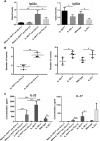
Altered immune response to Th‐1‐induced T. muris infection after conditional deletion of IL‐1R1. (A, B) ELISA measuring (A) specific T. muris IgG2 antibody in the blood and (B) worm burden counted from the colon and caecum, 21 days postinfection with low dose of T. muris eggs, of IL‐1R1fl/fl vav iCre‐, IL‐1R1fl/fl vav iCre+, IL‐1R1fl/fl, IL‐1R1−/−, IL‐22−/−, or wild‐type mice. (C) The blood levels of IL‐17 and IL‐22 from isolated MLN cells obtained from uninfected (naïve) or infected IL‐1R1fl/fl vav iCre‐, IL‐1R1fl/fl vav iCre+, IL‐1R1fl/fl, IL‐1R1−/−, IL‐22−/− or wild‐type mice was determined by ELISA. The data are presented as a mean ± SEM from three or four experiments carried out separately (n = 4 per experimental group except for left graphs in 3A and 3B where n = 3 per experimental group), and were analyzed statistically using one‐way ANOVA, followed by a Tukey multiple comparison post hoc test. *p < 0.05, **p < 0.01, ***p < 0.001.

References

    1. Glaccum, M. B. , Stocking, K. L. , Charrier, K. , Smith, J. L. , Willis, C. R. , Maliszewski, C. , Livingston, D. J. et al, Phenotypic and functional characterization of mice that lack the type I receptor for IL‐1. J. Immunol. 1997. 159: 3364–3371. - PubMed
    1. Labow, M. , Shuster, D. , Zetterstrom, M. , Nunes, P. , Terry, R. , Cullinan, E. B. , Bartfai, T. et al, Absence of IL‐1 signaling and reduced inflammatory response in IL‐1 type I receptor‐deficient mice. J. Immunol. 1997. 159: 2452–2461. - PubMed
    1. Humphreys, N. E. and Grencis, R. K. , IL‐1‐dependent, IL‐1R1‐independent resistance to gastrointestinal nematodes. Eur. J. Immunol. 2009. 39: 1036–1045. - PubMed
    1. Touzani, O. , Boutin, H. , LeFeuvre, R. , Parker, L. , Miller, A. , Luheshi, G. and Rothwell, N. , Interleukin‐1 influences ischemic brain damage in the mouse independently of the interleukin‐1 type I receptor. J. Neurosci. 2002. 22: 38–43. - PMC - PubMed
    1. Qian, J. , Zhu, L. , Li, Q. , Belevych, N. , Chen, Q. , Zhao, F. , Herness, S. and Quan, N. , Interleukin‐1R3 mediates interleukin‐1‐induced potassium current increase through fast activation of Akt kinase. Proc. Natl. Acad. Sci. USA 2012. 109: 12189–12194. - PMC - PubMed

Publication types