. 2015 Dec;38(4):420-32.
doi: 10.1590/S1415-475738420150019. Epub 2015 Nov 24.

Ionizing radiation-induced DNA injury and damage detection in patients with breast cancer

Affiliations

Ionizing radiation-induced DNA injury and damage detection in patients with breast cancer

Gissela Borrego-Soto et al. Genet Mol Biol. 2015 Dec.

Abstract

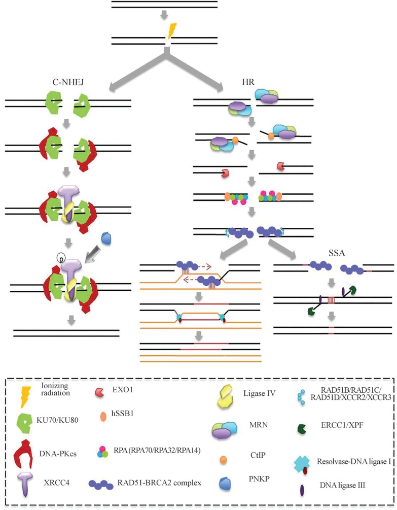
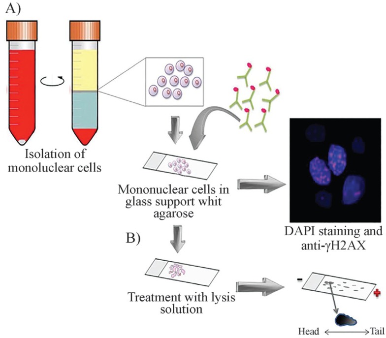
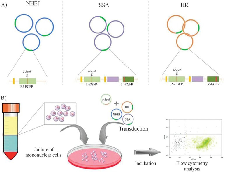
Breast cancer is the most common malignancy in women. Radiotherapy is frequently used in patients with breast cancer, but some patients may be more susceptible to ionizing radiation, and increased exposure to radiation sources may be associated to radiation adverse events. This susceptibility may be related to deficiencies in DNA repair mechanisms that are activated after cell-radiation, which causes DNA damage, particularly DNA double strand breaks. Some of these genetic susceptibilities in DNA-repair mechanisms are implicated in the etiology of hereditary breast/ovarian cancer (pathologic mutations in the BRCA 1 and 2 genes), but other less penetrant variants in genes involved in sporadic breast cancer have been described. These same genetic susceptibilities may be involved in negative radiotherapeutic outcomes. For these reasons, it is necessary to implement methods for detecting patients who are susceptible to radiotherapy-related adverse events. This review discusses mechanisms of DNA damage and repair, genes related to these functions, and the diagnosis methods designed and under research for detection of breast cancer patients with increased radiosensitivity.


Figures

Figure 1
Figure 1. DSB repair pathways. In NHEJ, the KU70/KU80 heterodimer binds to the DSB, protects it from degradation by exonucleases, and acts as a repressor of HR. The KU70/80 heterodimer recruits and activates the DNA-PKcs and KU70 interacts with XRCC4. Then, the DNA ligase IV interacts with the KU heterodimer to ligate the DNA ends. If required for ligation, PNKP binds to phosphorylated XRCC4 to process the DNA ends. In the HR pathway the MRN complex is recruited at the DSB ends and CtIP binds to the MRN complex activating an exonuclease activity which creates single strand segments at the borders of the DSB that are extended by the EXO1 3′- 5′ exonuclease. Then, hSSB1 binds to free ends and RPA (an heterometic complex formed by RPA70, RPA32 and RPA14) protects against degradation. RPA is replaced by RAD51-BRCA2. RAD51 nucleoprotein searches for and invades the homologues sequences, from sister chromatid, to form a Holliday junction. The sister chromatids are joined by cohesin proteins to facilitate the interconnection of the DSB to the homologous recombination. Subsequently, RAD51 is removed leaving a free 3′-OH and DNA is synthesized by the DNA polymerase δ using the homologous chromatid as a template. Resolvase enzymes solve the Holliday junction and the DNA ends are joined by DNA ligase I. The SSA pathway is not conservative and depends on the presence of repeated sequences flanking the DSB. In this mechanism, the MRN complex joined to CtIP cleaves the 5′-end of one strand of DNA to expose microhomology sequences. Homologous sequences are aligned, while nonaligned regions are removed by the ERCC1/XPF nucleases. Then, DNA ends are joined by DNA ligase III.
Figure 2
Figure 2. General assays for detecting DNA damage (A) Immunohistochemistry with antibodies directed against γ-H2AX: peripheral blood mononuclear cells are isolated, nuclei are stained with DAPI and with antibodies directed at γ-stained H2AX and visualized under fluorescent microscopy. (B) Comet assay: the comet assay is also performed on mononuclear cells. The cells are embedded in agarose on a thin glass slide, cells are lysed and incubated in an alkaline solution. Subsequently, DNA fragments are separated by electrophoresis and stained with ethidium bromide. The comet-like image is viewed under a fluorescence microscope. The length of the comet tail indicates the frequency of DNA breaks
Figure 3
Figure 3. Specific assays for detecting DNA damage (A) The EJ-EGFP plasmids contains a mutated version of the EGFP gene (green light bar) created by inserting a restriction site for the meganuclease I-SceI flanked by a 5 bp microhomology sites (black arrows); this plasmid was designed to be repaired by NHEJ. The Δ-EGFP/3’EGFP and Δ-EGFP/5’EGFP plasmids contain an array of an EGFP mutated gene containing an I-SceI site (green light bar) followed by a spacer (purple bar) and EGFP gene versions truncated at their flanking 3’ and 5’ ends, respectively (dark green bars) which allow the reconstitution of the wild-type version of the marker gene by SSA and HR, respectively. (B) Analysis of DSB repair: The assay is performed in three cultures of peripheral blood lymphocytes (PBLs), transduced separately with each of the plasmid versions designed for discrimination of SSA, NHEJ and HR. The cultures are co-transduced with an additional plasmid expressing the I-SceI enzyme. After generating DBS in the target plasmids by the expressed restriction enzyme, DNA repair in PBLs repair by each of the different DNA repair pathway may be monitored by restoration of the wild-type version of EGFP 24 h after transduction by measuring EGFP florescence by flow cytometry.

References

    1. Abraham ZH, Symonds N. Purification of overexpressed gam gene protein from bacteriophage Mu by denaturation-renaturation techniques and a study of its DNA-binding properties. Biochem J. 1990;269:679–684. - PMC - PubMed
    1. Alapetite C, Thirion P, De la Rochefordie A, Cosset JM, Moustacchi E. Analysis by alkaline comet assay of cancer patients with severe reactions to radiotherapy: Defective rejoining of radioinduced dna strand breaks in lymphocytes of breast cancer patients. Int J Cancer. 1999;90:83–90. - PubMed
    1. Aparicio T, Baer R, Gautier J. DNA double-strand break repair pathway choice and cancer. DNA Repair. 2014;19:169–175. - PMC - PubMed
    1. Azqueta A, Slyskova J, Langie SAS, Gaivão ION, Collins A. Comet assay to measure DNA repair: Approach and applications. Front Genet. 2014;5:1–8. - PMC - PubMed
    1. Barnett GC, West CML, Dunning AM, Elliott RM, Coles CE, Pharoah PDP, Burnet NG. Normal tissue reactions to radiotherapy: Towards tailoring treatment dose by genotype. Nat Rev Cancer. 2009;9:134–142. - PMC - PubMed