Review
. 2016 Jan;13(1):147-62.
doi: 10.1007/s13311-015-0410-6.

Antibody-Mediated Autoimmune Encephalopathies and Immunotherapies

Affiliations
Review

Antibody-Mediated Autoimmune Encephalopathies and Immunotherapies

Matteo Gastaldi et al. Neurotherapeutics. 2016 Jan.

Abstract

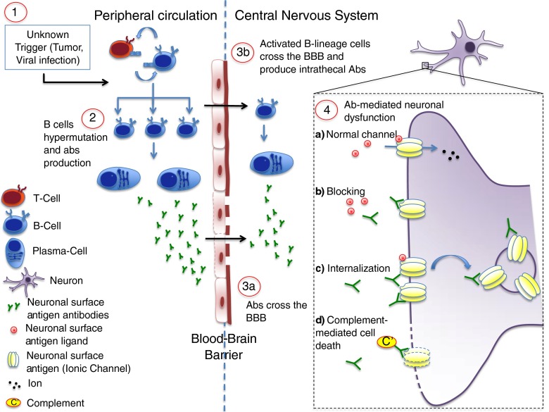
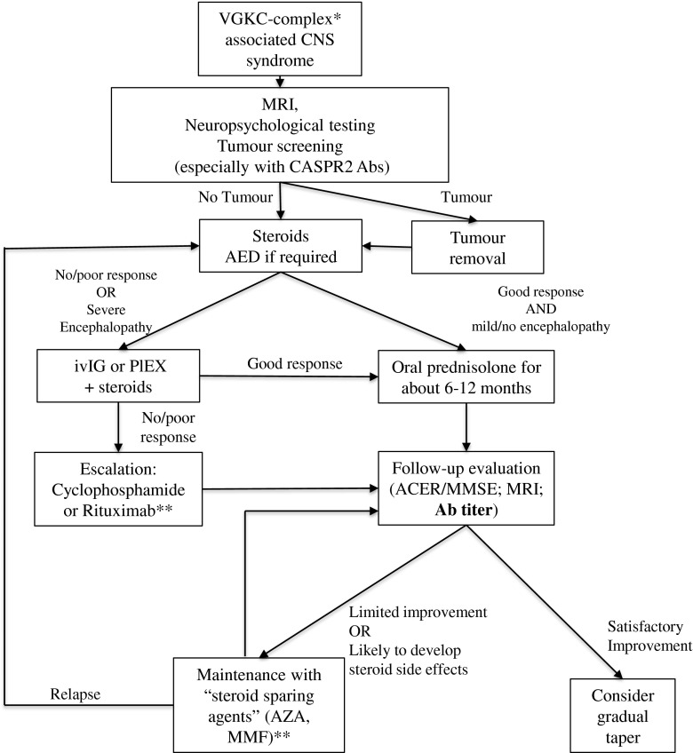
Over the last 15 years it has become clear that rare but highly recognizable diseases of the central nervous system (CNS), including newly identified forms of limbic encephalitis and other encephalopathies, are likely to be mediated by antibodies (Abs) to CNS proteins. The Abs are directed against membrane receptors and ion channel-associated proteins that are expressed on the surface of neurons in the CNS, such as N-methyl D-aspartate receptors and leucine-rich, glioma inactivated 1 protein and contactin-associated protein like 2, that are associated with voltage-gated potassium channels. The diseases are not invariably cancer-related and are therefore different from the classical paraneoplastic neurological diseases that are associated with, but not caused by, Abs to intracellular proteins. Most importantly, the new antibody-associated diseases almost invariably respond to immunotherapies with considerable and sometimes complete recovery, and there is convincing evidence of their pathogenicity in the relatively limited studies performed so far. Treatments include first-line steroids, intravenous immunoglobulins, and plasma exchange, and second-line rituximab and cyclophosphamide, followed in many cases by steroid-sparing agents in the long-term. This review focuses mainly on N-methyl D-aspartate receptor- and voltage-gated potassium channel complex-related Abs in adults, the clinical phenotypes, and treatment responses. Pediatric cases are referred to but not reviewed in detail. As there have been very few prospective studies, the conclusions regarding immunotherapies are based on retrospective studies.

Keywords: Autoantibodies; Autoimmune encephalopathy; Immunosuppressive treatment; N-methyl D-aspartate receptor; Voltage-gated potassium channel-complex.


Figures

Fig. 1
Fig. 1
Potential pathogenic mechanisms in antibody (Ab)-mediated autoimmune encephalopathy. The pathogenesis of Ab-mediated encephalopathies is still unclear. Several potential triggers have been proposed as the first determinant of an aberrant activation of the immune system (1). In N-methyl-D-aspartate encephalitis it is well recognized that a tumor (mainly an ovarian teratoma) or a herpetic infection can precede the onset of the disease, but in the majority of cases the trigger remains unknown. In the peripheral circulation B lymphocytes, after interaction with T-helper lymphocytes, become activated and undergo somatic hypermutation and differentiation, starting the auto-Ab production (2). Abs against neuronal surface Ag may subsequently reach the central nervous system by crossing the blood–brain barrier (BBB) at sites of increased permeability (3a). It is also likely that activated B-lineage cells are able to cross the BBB actively and undergo the same differentiation process within the central nervous system, contributing to the intrathecal pool of auto-Abs (3b). When the Abs reach their target, the normal function of the surface Ag (usually a ionic channel; 4a) can be altered by different mechanisms. The Abs may prevent the binding of the channel ligand (blocking; 4b); some Abs cause cross-linking and internalization of receptors and thus depletion from the cell surface (4c); finally, Abs may activate the complement cascade and induce neuronal death (4d)
Fig. 2
Fig. 2
Live and fixed cell-based assay (CBA) for the detection of neuronal surface antigen (Ag) Abs. The CBA is a technique that allows identification of Abs whilst preserving the tertiary structure of the antigen. Live human embryonic kidney (HEK) cells are transfected using plasmids that contain DNA coding for the antigenic target (1); transfected cells express the Ag mainly (but not exclusively) on their surface (2); cells can either be stained live (a), or be fixed and permeabilized (b), and are subsequently incubated with patient serum or cerebrospinal fluid (3). Specific Abs in the serum or cerebrospinal fluid will bind the expressed antigenic target; note that when cells are alive specific Abs are able to bind only antigenic targets expressed on the cell surface (a), whilst when cells are fixed/permeabilized intracellular Ags can be reached (b). Cells are then incubated with a secondary fluorescent Ab that recognizes human IgG (4) and the presence of fluorescent antihuman IgG is detected with a fluorescent microscope (5)
Fig. 3
Fig. 3
Management of voltage-gated potassium channel (VGKC) complex-associated disease. The algorithm describes a management approach to patients with central nervous system (CNS) syndromes and the presence in serum or cerebrospinal fluid of Abs directed against the VGKC complex, and it is intended as a general indication. Single patients could need a tailored approach based on the clinical phenotype *Mainly leucine-rich glioma inactivated 1 and contactin-associated protein like 2 (CASPR2) **The efficacy of different immunosuppressants has never been tested systematically in autoimmune encephalitis, and previous evidence suggests, for example, a better efficacy of mycophenolate mofetil (MMF) than cyclophosphamide in other autoimmune diseases [102]. Hence, the division between “escalating” and “steroid-sparing” drugs is not clear-cut and different strategies could be applied in different clinical settings MRI = magnetic resonance imaging; AED = antiepileptic drugs; IVIg = intravenous immunoglobulin; PLEX = plasma exchange; ACER = Addenbrooke’s Cognitive Examination Revised; MMSE = Mini-mental State Examination; AZA = azathioprine

Similar articles

Cited by

References

    1. Lancaster E, Dalmau J. Neuronal autoantigens—pathogenesis, associated disorders and antibody testing. Nat Rev Neurol. 2012;8:380–390. doi: 10.1038/nrneurol.2012.99. - DOI - PMC - PubMed
    1. Vincent A, Bien CG, Irani SR, Waters P. Autoantibodies associated with diseases of the CNS: new developments and future challenges. Lancet Neurol. 2011;10:759–772. doi: 10.1016/S1474-4422(11)70096-5. - DOI - PubMed
    1. Granerod J, Ambrose HE, Davies NWS, et al. Causes of encephalitis and differences in their clinical presentations in England: a multicentre, population-based prospective study. Lancet Infect Dis. 2010;10:835–844. doi: 10.1016/S1473-3099(10)70222-X. - DOI - PubMed
    1. Vitaliani R, Mason W, Ances B, Zwerdling T, Jiang Z, Dalmau J. Paraneoplastic encephalitis, psychiatric symptoms, and hypoventilation in ovarian teratoma. Ann Neurol. 2005;58:594–604. doi: 10.1002/ana.20614. - DOI - PMC - PubMed
    1. Dalmau J, Tuzun E, Wu HY, et al. Paraneoplastic anti-N-methyl-D-aspartate receptor encephalitis associated with ovarian teratoma. Ann Neurol. 2007;61:25–36. doi: 10.1002/ana.21050. - DOI - PMC - PubMed

MeSH terms

Substances