

Tuberculous Conjunctivitis in an Anophthalmic Socket
- PMID: 26692731
- PMCID: PMC4660546
- DOI: 10.4103/0974-9233.167828
Tuberculous Conjunctivitis in an Anophthalmic Socket
Abstract
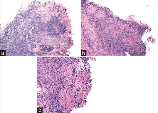
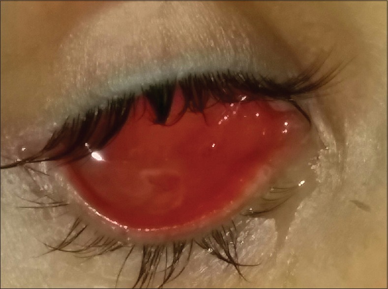
Tuberculous (TB) conjunctivitis was not an uncommon condition before the early 20(th) century but is currently a rare occurrence, especially in the developed countries. We report a 27-year-old Saudi female who underwent enucleation of the right eye at the age of 20 following a penetrating eye injury. She had a history of miliary TB that was treated at the age of 22. She was presented with chronic purulent discharge from her right an anophthalmic socket for 2 months. Cultures for bacteria and fungi were sterile. There was no response to empirical topical antibiotics and steroids. Direct microscopic examination of conjunctival scrapings with the Ziehl-Neelsen staining revealed no microorganisms. Histopathological examination revealed epithelioid granulomas. Polymerase chain reaction was negative for Mycobacterium tuberculosis DNA. TB conjunctivitis was suspected from the history of miliary TB and presence of epithelioid granulomas. The definitive diagnosis was made after prompt resolution of the ocular signs with no recurrence only after systemic anti-TB therapy.
Keywords: Anophthalmic Socket; Anti-tuberculous Therapy; Enucleation; Miliary Tuberculosis; Tuberculous Conjunctivitis.
Figures
Similar articles
-
Mycobacterium tuberculosis presenting as chronic red eye.Cornea. 2006 Oct;25(9):1118-20. doi: 10.1097/01.ico.0000240097.99536.82. Cornea. 2006. PMID: 17133069
-
Painful Red Eye in a Woman in Her 70s.JAMA Ophthalmol. 2016 Feb;134(2):233-4. doi: 10.1001/jamaophthalmol.2015.2152. JAMA Ophthalmol. 2016. PMID: 26720132 No abstract available.
-
Unilateral tuberculous conjunctivitis with tarsal necrosis.Arch Ophthalmol. 2003 Oct;121(10):1475-8. doi: 10.1001/archopht.121.10.1475. Arch Ophthalmol. 2003. PMID: 14557188 No abstract available.
-
Tuberculous subcutaneous abscesses developing during miliary tuberculosis therapy.Scand J Infect Dis. 2000;32(1):37-40. doi: 10.1080/00365540050164191. Scand J Infect Dis. 2000. PMID: 10716075 Review.
-
[Ocular tuberculosis].Rev Chilena Infectol. 2007 Aug;24(4):284-95. doi: 10.4067/s0716-10182007000400004. Epub 2007 Aug 20. Rev Chilena Infectol. 2007. PMID: 17728915 Review. Spanish.
References
-
- Gupta V, Gupta A, Rao NA. Intraocular tuberculosis NA.n update. Surv Ophthalmol. 2007;52:561–87. - PubMed
-
- Bodaghi B, Le Hoang P. Ocular tuberculosis. Curr Opin Ophthalmol. 2000;11:443–8. - PubMed
-
- Eyre JW. Tuberculosis of the conjunctiva: Its etiology, pathology, and diagnosis. Lancet. 1912;1:1319–28.
-
- Biswas J, Badrinath SS. Ocular morbidity in patients with active systemic tuberculosis. Int Ophthalmol. 1995-1996;19:293–8. - PubMed
-
- Salas D, Murthy S, Champ C, Hawksworth N. Primary tuberculosis of the conjunctiva. Eye (Lond) 2001;15(Pt 5):674–6. - PubMed
Publication types
MeSH terms
Substances
LinkOut - more resources
Full Text Sources
Other Literature Sources
