. 2015 Dec 12:15:116.
doi: 10.1186/s12935-015-0269-y. eCollection 2015.

Arsenic trioxide reduces chemo-resistance to 5-fluorouracil and cisplatin in HBx-HepG2 cells via complex mechanisms

Affiliations

Arsenic trioxide reduces chemo-resistance to 5-fluorouracil and cisplatin in HBx-HepG2 cells via complex mechanisms

Guifang Yu et al. Cancer Cell Int. .

Abstract

Background: Multidrug resistance is one of the major reasons chemotherapy-based treatments failed in hepatitis B virus (HBV) related hepatocellular carcinoma (HCC). Hypoxia is generally associated with tumor chemo-resistance. The aim of the study was to investigate the effect of Arsenic trioxide (As2O3) on the hypoxia-induced chemo-resistance to 5-FU or cisplatin and explored its underlying mechanism in the HBx-HepG2 cells.

Methods: MTT assay was used to examine the cell viability. Mitochondrial membrane potential (MMP) and cell cycle was examined by flow cytometry. qRT-PCR was employed to observe the mRNA expression level; and western blot assay was used to determine the protein expression level.

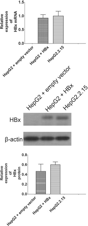
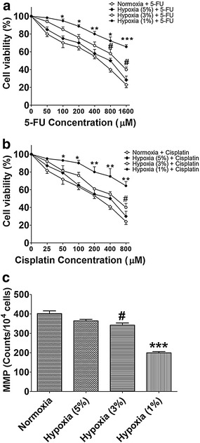
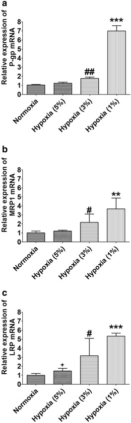
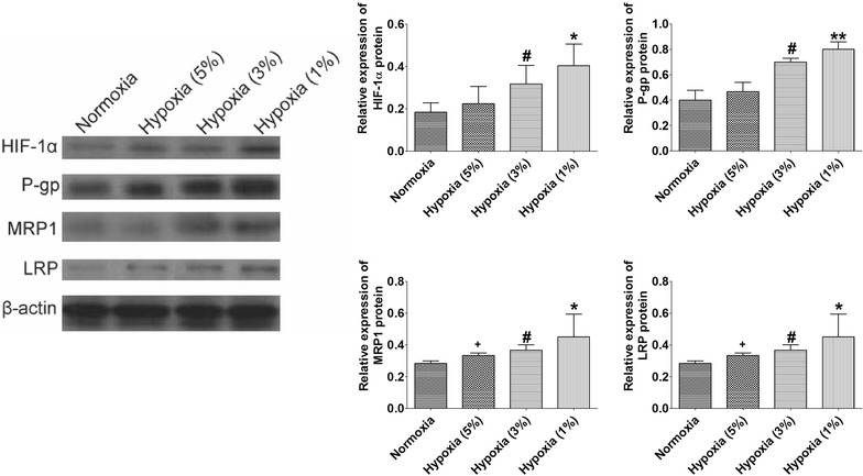
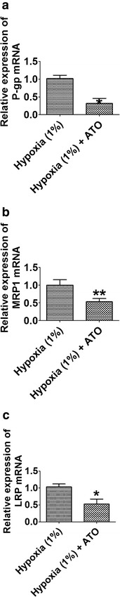
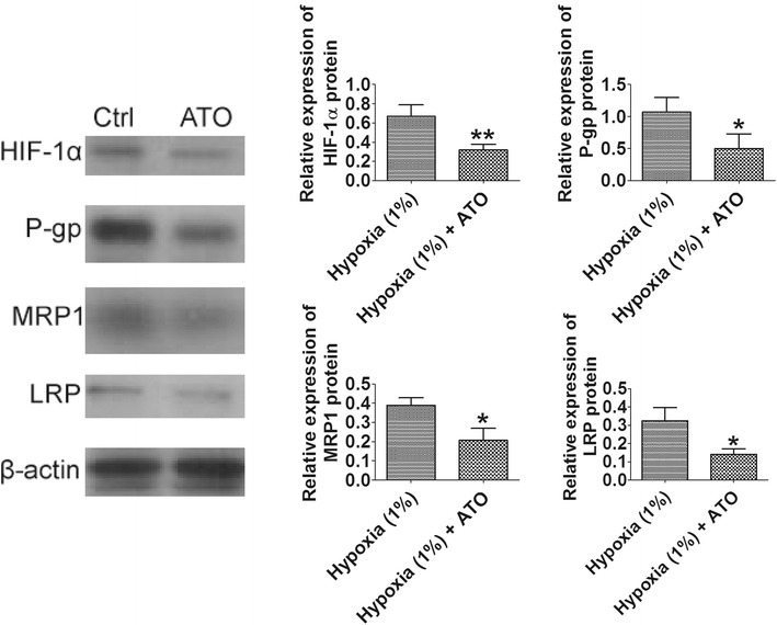
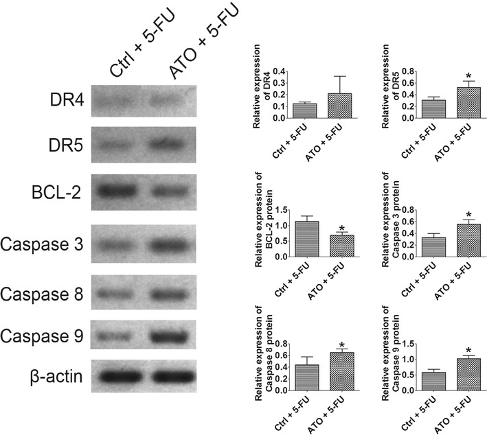
Results: Our results showed that transfection of HBx plasmid established the HBx-HepG2 cells expressing HBx, and the expression of HBx was confirmed by qRT-PCR and western blot. Exposure of HBx-HepG2 cells to hypoxia (5 % O2, 3 % O2, 1 % O2) for 48 h increased the chemo-resistance to 5-fluorouracil (5-FU) (50-1600 µM) and cisplatin (25-800 µM), reduced MMP, and caused the cell cycle arrest at G0/G1 phase in a concentration-dependent manner. Hypoxia also concentration-dependently (5 % O2, 3 % O2, 1 % O2) reduced mRNA expression level of P-glycoprotein (P-gp), multidrug resistance protein (MRP1), lung resistance protein (LRP), and decreased the protein expression level of hypoxia-inducible factor-1α (HIF-1α), P-gp MRP1, and LRP. Following pretreatment with As2O3 at a non-cytotoxic concentration re-sensitized the hypoxia (1 % O2)-induced chemo-resistance to 5-FU and cisplatin in HBx-HepG2 cells. As2O3 pretreatment also prevented MMP reduction and G0/G1 arrest induced by hypoxia. Meanwhile, As2O3 antagonized increase of HIF-1α protein induced by hypoxia, and it also suppresses the increase in expression levels of P-gp, MRP1, and LRP mRNA and proteins. In addition, As2O3 in combination with 5-FU treatment caused up-regulation of DR5, caspase 3, caspase 8, and caspase 9, and down-regulation of BCL-2, but had no effect of DR4.

Conclusions: Our results may suggest that As2O3 re-sensitizes hypoxia-induced chemo-resistance in HBx-HepG2 via complex pathways, and As2O3 may be a potential agent that given in combination with other anti-drugs for the treatment of HBV related HCC, which is resistant to chemotherapy.

Keywords: 5-FU; Chemo-resistance; Cisplatin; HBV; HBx-HepG2; HCC; HIF-1α; LRP; MRP1; P-gp.


Figures

Fig. 1
Fig. 1
Establishment of HepG2-HBx cell lines. qRT-PCR and western blot were used to evaluate the expression levels of HBx mRNA and protein in empty vector-transfected HepG2 cells, HBx-transfected HepG2 cells and HepG2.2.15 cells. All data were expressed as mean ± s.e.m., n = 4
Fig. 2
Fig. 2
Effect of hypoxia on the cell viability of HBX-HepG2 cells treated with chemotherapeutic agents and on the mitochondrial membrane potential (MMP) in HBX-HepG2 cells. MTT assay was conducted on HBX-HepG2 cells pre-incubated for 48 h in normoxic (21 % O2) or hypoxic (5 % O2, 3 % O2, or 1 % O2) conditions and subsequently treated with a 5-FU (50–1600 µM) or b Cisplatin (25–800 µM); c flow-cytometry was conducted on HBx-HepG2 cells pre-incubated for 48 h in normoxic (21 % O2) or hypoxic (5 % O2, 3 % O2, or 1 % O2) conditions to measure the MMP. All data were expressed as mean ± s.e.m., n = 4; significant differences between hypoxia (1 % O2) group and normoxia group were indicated as *P < 0.05, **P < 0.01, ***P < 0.001; significantly differences between hypoxia (3 % O2) group and normoxia group were indicated as # P < 0.05 (One-way ANOVA followed by Bonferroni’s multiple comparison tests)
Fig. 3
Fig. 3
Effect of hypoxia on expression levels of P-gp, MRP1 and LRP mRNA inHBX-HepG2 cells. qRT-PCR assay was conducted on HBx-HepG2 cells pre-incubated for 48 h in normoxic (21 % O2) or hypoxic (5 % O2, 3 % O2, or 1 % O2) to measure the expression levels of a P-gp, b MRP1 and c LRP mRNA. All data were expressed as mean ± s.e.m., n = 4; significant differences between hypoxia (1 % O2) group and normoxia group were indicated as **P < 0.01, ***P < 0.001; significant differences between hypoxia (3 % O2) group and normoxia group were indicated as # P < 0.05; ## P < 0.01 (One-way ANOVA followed by Bonferroni’s multiple comparison tests)
Fig. 4
Fig. 4
Effect of hypoxia on expression levels of HIF-1α, P-gp, MRP1 and LRP protein in HBx-HepG2 cells. Western blot assay was conducted on HBX-HepG2 cells pre-incubated for 48 h in normoxic (21 % O2) or hypoxic (5 % O2, 3 % O2, or 1 % O2) conditions to measure the expression levels of HIF-1α, P-gp, MRP1 and LRP protein, band densities were normalized to β-actin. All data were expressed as mean ± s.e.m., n = 4; significant differences between hypoxia (1 % O2) group and normoxia group were indicated as *P < 0.05, **P < 0.01; significantly differences between hypoxia (3 % O2) group and normoxia group were indicated as # P < 0.05 (One-way ANOVA followed by Bonferroni’s multiple comparison tests)
Fig. 5
Fig. 5
Effect of As2O3 (1 µM) on chemo-sensitivity, MMP, and cell cycle of HBx-HepG2 cells pretreated with hypoxia (1 % O2). MTT assay was used to evaluate the effect of As2O3 on chemo-sensitivity to a 5-FU (50–1600 µM) or b cisplatin (25–800 µM) in HBx-HepG2 cells pretreated with 48 h hypoxia (1 % O2); c flow-cytometry was used to examine the effect of As2O3 on MMP in HBx-HepG2 cells pretreated with 48 h hypoxia (1 % O2); d flow-cytometry was used to examine the effect of As2O3 on MMP in HBx-HepG2 cells pretreated with 48 h hypoxia (1 % O2). All data were expressed as mean ± s.e.m., n = 4; ATO = As2O3; significant differences between hypoxia (1 % O2) group and hypoxia (1 % O2) + ATO group were indicated as *P < 0.05, **P < 0.01, ***P < 0.001 (unpaired t-test)
Fig. 6
Fig. 6
Effect of As2O3 (1 µM) on the expression levels of P-gp, MRP1 and LRP mRNA inHBx-HepG2 cells pretreated with hypoxia (1 % O2). qRT-PCR assay was used to examine the effect of As2O3 on the expression levels of a P-gp, b MRP1 and c LRP mRNA in HBx-HepG2 cells pretreated with hypoxia (1 % O2). All data were expressed as mean ± s.e.m., n = 4; significantly differences between hypoxia (1 % O2) group and hypoxia (1 % O2) + ATO group were indicated as *P < 0.05, **P < 0.01 (unpaired t-test)
Fig. 7
Fig. 7
Effect of As2O3 (1 µM) on the expression levels of P-gp, MRP1 and LRP protein in HBx-HepG2 cells pretreated with hypoxia (1 % O2). Western blot assay was used to examine the effect of As2O3 on the expression levels of P-gp, MRP1 and LRP protein in HBx-HepG2 cells pretreated with hypoxia (1 % O2), band densities were normalized to β-actin. All data were expressed as mean ± s.e.m., n = 4; significant differences between hypoxia (1 % O2) group and hypoxia (1 % O2) + ATO group were indicated as *P < 0.05, **P < 0.01 (unpaired t-test)
Fig. 8
Fig. 8
Effect of As2O3 (1 µM) + 5-FU (400 µM) on the expression levels of apoptotic-related factors protein in HBx-HepG2 cells pretreated with hypoxia (1 % O2). Western blot assay was used to examine the effect of As2O3 + 5-FU on the expression levels of DR4, DR5, BCL-2, caspase 3, caspase 8 and caspase 9 protein in HBx-HepG2 cells pretreated with hypoxia (1 % O2), band densities were normalized to β-actin. All data were expressed as mean ± s.e.m., n = 4; significant differences between Ctrl + 5-FU group and ATO + 5-FU group were indicated as *P < 0.05 (unpaired t-test)

References

    1. Sinn DH, Lee J, Goo J, Kim K, Gwak GY, Paik YH, Choi MS, Lee JH, Koh KC, Yoo BC, et al. Hepatocellular carcinoma risk in chronic hepatitis B virus-infected compensated cirrhosis patients with low viral load. Baltimore: Hepatology; 2015. - PubMed
    1. Liu XY, Tang SH, Wu SL, Luo YH, Cao MR, Zhou HK, Jiang XW, Shu JC, Bie CQ, Huang SM, et al. Epigenetic modulation of insulin-like growth factor-II overexpression by hepatitis B virus X protein in hepatocellular carcinoma. Am J cancer Res. 2015;5(3):956–978. - PMC - PubMed
    1. Yin D, Huang P, Zhuang B, Zhang H, Yan H, Xiao Z, Li W, Zhang J, Tang Q, Hu K, et al. Hepatitis B virus X protein (HBx) is responsible for resistance to targeted therapies in hepatocellular carcinoma: ex vivo culture evidence. Clin Cancer Res. 2015;21(49):4420–4430. - PubMed
    1. Koike K. Hepatocarcinogenesis in hepatitis viral infection: lessons from transgenic mouse studies. J Gastroenterol. 2002;37(Suppl 13):55–64. doi: 10.1007/BF02990101. - DOI - PubMed
    1. Chami M, Ferrari D, Nicotera P, Paterlini-Brechot P, Rizzuto R. Caspase-dependent alterations of Ca2+ signaling in the induction of apoptosis by hepatitis B virus X protein. J Biol Chem. 2003;278(34):31745–31755. doi: 10.1074/jbc.M304202200. - DOI - PubMed