

Protective effects of icariin on cisplatin-induced acute renal injury in mice
- PMID: 26692955
- PMCID: PMC4656788
Protective effects of icariin on cisplatin-induced acute renal injury in mice
Abstract
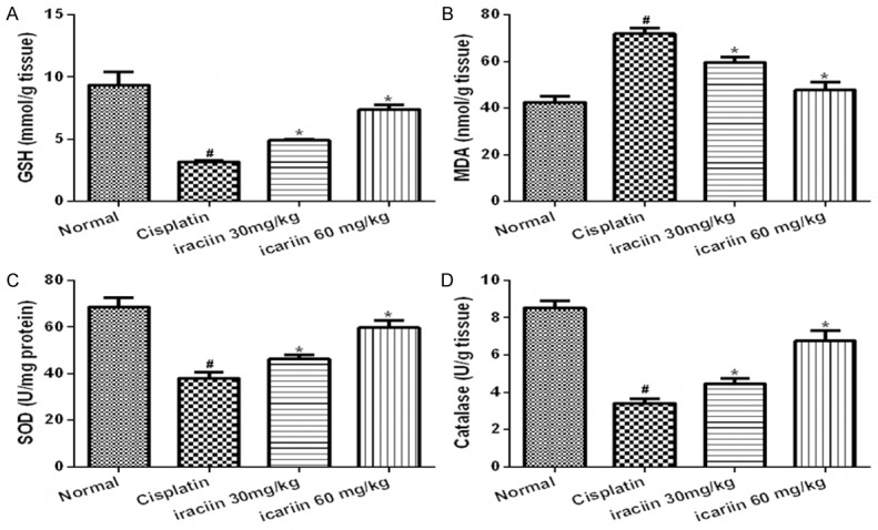
Cisplatin chemotherapy often causes acute kidney injury in cancer patients. Icariin is a bioactive flavonoid, which has renal protection and anti-inflammation effects. This study investigated the mechanism underlying the attenuation of cisplatin-induced renal injury by icariin. BALB/c mice were treated with cisplatin (15 mg/kg) with or without treatment with icariin (30 or 60 mg/kg for 5 days). Renal function, histological changes, degree of oxidative stress and tubular apoptosis were examined. The effects of icariin on cisplatin-induced expression of renal TNF-α, NF-κB, cleaved caspase-3 and Bcl-2 family proteins were evaluated. Treatment of mice with cisplatin resulted in renal damage, showing an increase in blood urea nitrogen and creatinine levels, tubular damage, oxidative stress and apoptosis. These renal changes could be significantly improved by icariin treatment, especially in high dose of icariin group. Examination of molecules involving inflammation and apoptosis of the kidney revealed that treatment of icariin reduced expression of TNF-α, NF-κB, cleaved caspase-3, and Bax, increased the expression of BCL-2. These results indicate that icariin ameliorates the cisplatin-mediated nephrotoxicity via improving renal oxidant status, consequent NF-κB activation and inflammation cascade and apoptosis, and the following disturbed expression of apoptosis related proteins.
Keywords: Acute kidney injury; apoptosis; cisplatin; icariin; inflammation.
Figures


Similar articles
-
Icariin Improves Sepsis-Induced Mortality and Acute Kidney Injury.Pharmacology. 2018;102(3-4):196-205. doi: 10.1159/000487955. Epub 2018 Aug 10. Pharmacology. 2018. PMID: 30099451
-
Icariin ameliorates cisplatin-induced cytotoxicity in human embryonic kidney 293 cells by suppressing ROS-mediated PI3K/Akt pathway.Biomed Pharmacother. 2019 Jan;109:2309-2317. doi: 10.1016/j.biopha.2018.11.108. Epub 2018 Nov 29. Biomed Pharmacother. 2019. PMID: 30551489
-
Protective Effects of Luteolin on Lipopolysaccharide-Induced Acute Renal Injury in Mice.Med Sci Monit. 2016 Dec 28;22:5173-5180. doi: 10.12659/msm.898177. Med Sci Monit. 2016. PMID: 28029146 Free PMC article.
-
Dexmedetomidine protects against cisplatin-induced acute kidney injury in mice through regulating apoptosis and inflammation.Inflamm Res. 2017 May;66(5):399-411. doi: 10.1007/s00011-017-1023-9. Epub 2017 Feb 21. Inflamm Res. 2017. PMID: 28224201
-
Icariin: A Promising Natural Product in Biomedicine and Tissue Engineering.J Funct Biomater. 2023 Jan 12;14(1):44. doi: 10.3390/jfb14010044. J Funct Biomater. 2023. PMID: 36662090 Free PMC article. Review.
Cited by
-
Farrerol Attenuates Cisplatin-Induced Nephrotoxicity by Inhibiting the Reactive Oxygen Species-Mediated Oxidation, Inflammation, and Apoptotic Signaling Pathways.Front Physiol. 2019 Nov 26;10:1419. doi: 10.3389/fphys.2019.01419. eCollection 2019. Front Physiol. 2019. PMID: 31849693 Free PMC article.
-
Rapamycin induces autophagy to alleviate acute kidney injury following cerebral ischemia and reperfusion via the mTORC1/ATG13/ULK1 signaling pathway.Mol Med Rep. 2018 Dec;18(6):5445-5454. doi: 10.3892/mmr.2018.9586. Epub 2018 Oct 24. Mol Med Rep. 2018. PMID: 30365078 Free PMC article.
-
Beneficial Effects of Bioactive Compounds in Mulberry Fruits against Cisplatin-Induced Nephrotoxicity.Int J Mol Sci. 2018 Apr 9;19(4):1117. doi: 10.3390/ijms19041117. Int J Mol Sci. 2018. PMID: 29642519 Free PMC article.
-
Icariin improves Fanconi anemia hematopoietic stem cell function through SIRT6-mediated NF-kappa B inhibition.Cell Cycle. 2018;17(3):367-376. doi: 10.1080/15384101.2018.1426413. Epub 2018 Feb 8. Cell Cycle. 2018. PMID: 29355456 Free PMC article.
-
Pro-Inflammatory Signalling PRRopels Cisplatin-Induced Toxicity.Int J Mol Sci. 2022 Jun 29;23(13):7227. doi: 10.3390/ijms23137227. Int J Mol Sci. 2022. PMID: 35806229 Free PMC article. Review.
References
-
- Yao X, Panichpisal K, Kurtzman N, Nugent K. Cisplatin nephrotoxicity: a review. Am J Med Sci. 2007;334:115–124. - PubMed
-
- Kuhlmann MK, Burkhardt G, Köhler H. Insights into potential cellular mechanisms of cisplatin nephrotoxicity and their clinical application. Nephrol Dial Transplant. 1997;12:2478–2480. - PubMed
-
- Camano S, Lazaro A, Moreno-Gordaliza E, Torres AM, de Lucas C, Humanes B, Lazaro JA, Gomez-Gomez MM, Bosca L, Tejedor A. Cilastatin attenuates cisplatin-induced proximal tubular cell damage. J Pharmacol Exp Ther. 2010;334:419–429. - PubMed
-
- Al-Majed AA, Sayed-Ahmed MM, Al-Yahya AA, Aleisa AM, Al-Rejaie SS, Al-Shabanah OA. Propionyl-L-carnitine prevents the progression of cisplatin-induced cardiomyopathy in a carnitine-depleted rat model. Pharmacol Res. 2006;53:278–286. - PubMed
LinkOut - more resources
Full Text Sources
Research Materials