. 2015:2015:182495.
doi: 10.1155/2015/182495. Epub 2015 Nov 26.

The Impact of Paeoniflorin on α-Synuclein Degradation Pathway

Affiliations

The Impact of Paeoniflorin on α-Synuclein Degradation Pathway

Zenglin Cai et al. Evid Based Complement Alternat Med. 2015.

Abstract

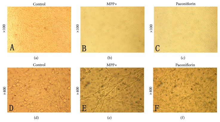
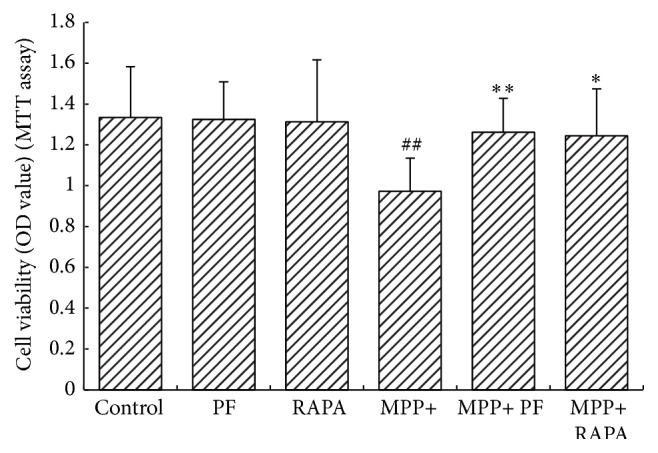
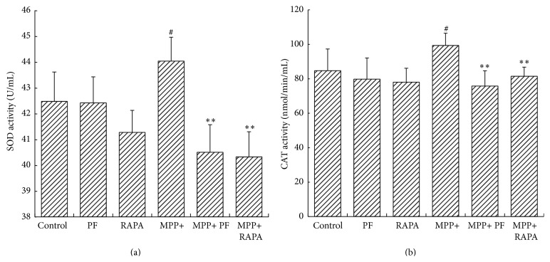
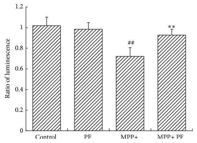
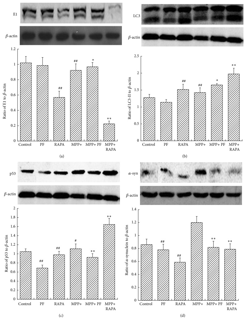
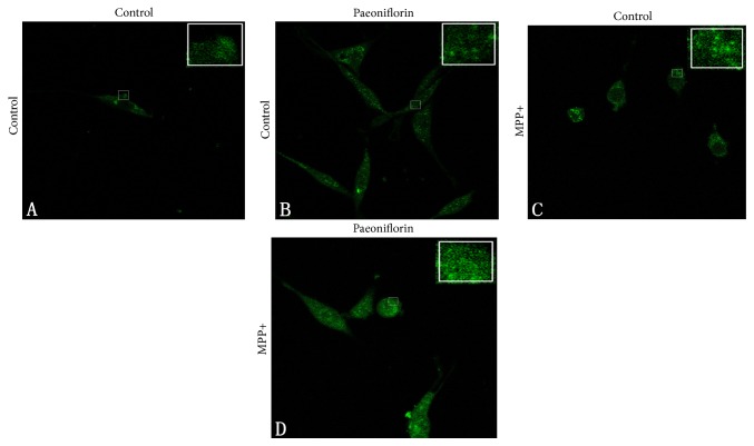
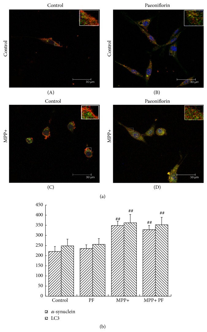
Paeoniflorin (PF) is the major active ingredient in the traditional Chinese medicine Radix. It plays a neuroprotective role by regulating autophagy and the ubiquitin-proteasome degradation pathway. In this study, we found PF significantly reduced cell damage caused by MPP+, returning cells to normal state. Cell viability significantly improved after 24 h exposure to RAPA and PF in the MPP+ group (all P < 0.01). CAT and SOD activities were significantly decreased after PF and RAPA treatment, compared with MPP+ (P < 0.001). In addition, MPP+ activated both LC3-II and E1; RAPA increased LC3-II but inhibited E1. PF significantly upregulated both LC3-II (autophagy) and E1 (ubiquitin-proteasome pathway) expression (P < 0.001), promoted degradation of α-synuclein, and reduced cell damage. We show MPP+ enhanced immunofluorescence signal of intracellular α-synuclein and LC3. Fluorescence intensity of α-synuclein decreased after PF treatment. In conclusion, these data show PF reversed the decline of proteasome activity caused by MPP+ and significantly upregulated both autophagy and ubiquitin-proteasome pathways, promoted the degradation of α-synuclein, and reduced cell damage. These findings suggest PF is a potential therapeutic medicine for neurodegenerative diseases.


Figures

Figure 1
Figure 1
Paeoniflorin shows a protective effect from injury by MPP+ under light microscopy. (a) Normal PC12 cells (×100). (b) MPP+ treated PC12 cells at 24 h (×100-fold). (c) Paeoniflorin treated cells 24 h after MPP+ treatment (×100). (d) Normal PC12 cells (×400). (e) MPP+ 24 h (×400). (f) Paeoniflorin treated cells 24 h after MPP+ treatment (×400).
Figure 2
Figure 2
MTT cell viability assay. Both PF and Rapamycin treatment significantly improved cell viability in MPP+ treated group (# P < 0.05, ## P < 0.01 versus control group; P < 0.05, ∗∗ P < 0.01 versus corresponding control group; mean ± SD, n ≥ 6).
Figure 3
Figure 3
Superoxide dismutase (SOD) and catalase (CAT) activity in cells. (a) Rapamycin and PF significantly decreased SOD activity after MPP+ treatment. (b) CAT activity decreased in MPP+ group (# P < 0.05, ## P < 0.01 versus control group; P < 0.05, ∗∗ P < 0.01 versus corresponding control group, mean ± SD, n ≥ 6).
Figure 4
Figure 4
Change in proteasome activity following MPP+ and PF treatment. Under normal circumstances, the impact of PF on proteasome activity was not statistically significant (P = 0.076). MPP+ inhibited the ubiquitin-proteasome activity (P = 0.002). Paeoniflorin reversed the decline of proteasome activity caused by MPP+ (P = 0.004) (## P < 0.01 MPP+ group versus control group; ∗∗ P < 0.01 versus MPP+ group; mean ± SD, n ≥ 6).
Figure 5
Figure 5
Rapamycin and PF impact on α-synuclein and its degradation pathway. Western blots (upper panel in (a), (b), (c), and (d)) and statistical analysis of optical density measurements (lower panel in (a), (b), (c), and (d)) in PC12 cells after treatment with MPP+, PF, and RAPA for (a) E1, (b) LC3-II, (c) p53, and (d) α-synuclein. In MPP+ group, RAPA increased LC3-II and inhibited E1. PF upregulated both LC3-II and E1 significantly. Values represent mean ± SEM (n = 5). # P < 0.05, ## P < 0.01 versus control group. P < 0.05, ∗∗ P < 0.01 versus corresponding control group.
Figure 6
Figure 6
Paeoniflorin and MPP+ treatment on α-synuclein aggregation: (A) normal condition; (B) Paeoniflorin treatment did not affect α-synuclein aggregation; (C) after MPP+ treatment, α-synuclein tends to accumulate and aggregates appeared in the cytoplasm; (D) but after MPP+ and then Paeoniflorin treatment, cytoplasmic α-synuclein aggregates significantly reduced.
Figure 7
Figure 7
α-synuclein and LC3 colocalization in PC12 cells. Immunostain of α-synuclein (green) and LC3 (red) (a) and statistical analysis of optical density measurements (b), under normal conditions (A) and Paeoniflorin treatment (B), did not affect α-synuclein and LC3 and their colocalization; after MPP+ treatment (C), the signals of α-synuclein and LC3 were increased and lost their colocalization (more obvious in the enlarged figures). LC3 mainly aggregated in the peripheral cytoplasm but α-synuclein was distributed throughout the cell body. After MPP+ treatment and Paeoniflorin (D), the signals of α-synuclein and LC3 were both decreased and colocalization remained evident. Scale bar: 30 um. Quantification of immunostain results showed Paeoniflorin treatment did not affect fluorescence intensity of α-synuclein and LC3 in normal growth state (P > 0.05) but decreased the fluorescence intensity of α-synuclein and LC3 in MPP+ group. Values represent mean ± SEM (n = 5). # P < 0.05, ## P < 0.01 versus control group.

References

    1. Vekrellis K., Xilouri M., Emmanouilidou E., Rideout H. J., Stefanis L. Pathological roles of alpha-synuclein in neurological disorders. The Lancet Neurology. 2011;10(11):1015–1025. doi: 10.1016/s1474-4422(11)70213-7. - DOI - PubMed
    1. Kalia L. V., Kalia S. K., McLean P. J., Lozano A. M., Lang A. E. α-Synuclein oligomers and clinical implications for parkinson disease. Annals of Neurology. 2013;73(2):155–169. doi: 10.1002/ana.23746. - DOI - PMC - PubMed
    1. Wang K., Zhu L., Zhu X., et al. Protective effect of paeoniflorin on Abeta25-35-induced SH-SY5Y cell injury by preventing mitochondrial dysfunction. Cellular and Molecular Neurobiology. 2014;34(2):227–234. doi: 10.1007/s10571-013-0006-9. - DOI - PMC - PubMed
    1. Lin B. Polyphenols and neuroprotection against ischemia and neurodegeneration. Mini-Reviews in Medicinal Chemistry. 2011;11(14):1222–1238. - PubMed
    1. Guo R.-B., Wang G.-F., Zhao A.-P., Gu J., Sun X.-L., Hu G. Paeoniflorin protects against ischemia-induced brain damages in rats via inhibiting MAPKs/NF-κB-mediated inflammatory responses. PLoS ONE. 2012;7(11) doi: 10.1371/journal.pone.0049701.e49701 - DOI - PMC - PubMed

LinkOut - more resources