Review
. 2015 Mar 1;2(1):R37-44.
doi: 10.1530/ERP-15-0005. Epub 2015 Mar 9.

Surgical management of left ventricular outflow obstruction in hypertrophic cardiomyopathy

Affiliations
Review

Surgical management of left ventricular outflow obstruction in hypertrophic cardiomyopathy

Neil Howell et al. Echo Res Pract. .

Abstract

Hypertrophic cardiomyopathy is the single most common form of inherited heart disease. Left ventricular outflow tract obstruction (LVOTO) is a recognised feature of this condition which arises when blood leaving the outflow tract is impeded by systolic anterior motion of the mitral valve. In an important minority of patients, breathlessness, chest pain and syncope may result and persist despite the use of medications. In suitable candidates, surgery may relieve obstruction and its associated symptoms, and normalise life expectancy. Refinements in surgical techniques have marked improvements in the understanding of mechanisms underlying LVOTO. In this review, we hope to provide the reader with an understanding of how contemporary surgical practice has developed, which patients should be considered for surgery, and what results are anticipated. The role echocardiography plays in this area is highlighted throughout.

Keywords: hypertrophic cardiomyopathy; left ventricular outflow tract obstruction; myectomy.


Figures

Figure 1
Figure 1
Scheme portraying how SAM evolves over systole (A, B and C) and why posteriorly directed mitral regurgitation (C) is seen. This image is licensed under an unspecified non-commercial open access CC licence, and is reproduced with permission from Elsevier, from Grigg LE, Wigle ED, Williams WG, Daniel LB & Rakowski H 1992 Transesophageal Doppler echocardiography in obstructive hypertrophic cardiomyopathy: clarification of pathophysiology and importance in intraoperative decision making. Journal of the American College of Cardiology 20 42–52 .
Figure 2
Figure 2
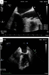
Systolic anterior motion of the mitral valve as seen by transoesophageal echocardiography in the mid-oesophageal long-axis (A) and five-chamber views (B). SAM–septal contact is seen in both. Measurements are used to describe septal anatomy are shown in (A). See main text for further details.
Figure 3
Figure 3
Assessment of LVOT obstruction using transoesophageal echocardiography from the deep transgastric position. The point of obstruction is denoted by the narrowest part of the jet seen on colour Doppler (A, arrows). By directing continous wave Doppler through the centre of the jet, a clear late peaking gradient can be attained (B). In this case the peak gradient was 91 mmHg.
Figure 4
Figure 4
A fibrous ridge causing subaortic obstruction as visualised using transoesophageal echocardiography (A, arrow). The minimal area during systole has been quantified by three-dimensional analysis (B).
Figure 5
Figure 5
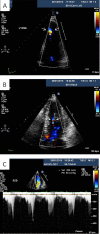
LV obstruction remote to the outflow tract assessed by transthoracic echocardiography: apical obstruction (A) and mid ventricular obstruction (B) identified by colour Doppler (arrows). In the patient in B, a peak gradient of 50 mmHg was derived (C).
Figure 6
Figure 6
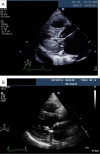
The effect of myectomy on LVOT anatomy: pre-myectomy in an individual who had previously undergone alcohol septal ablation (A). Severe mitral regurgitation due to endocarditis (not shown) had necessitated emergency surgery. Note how the chordate contact the hypertrophied septum (arrow). Post-operative images following mitral valve repair and myectomy (B) show how the LVOT is wider with chordae positioned well away from the ventricular septum. A mitral valve annuloplasty ring is also present.
Figure 7
Figure 7
Iatrogenic post-myectomy ventricular septal defect. Colour flow is seen crossing a ventricular septal defect (arrows) sited between the left ventricle (LV) and right ventricle (RV) in a modified four chamber view using transthoracic echocardiography.

References

    1. Nagueh SF Bierig SM Budoff MJ Desai M Dilsizian V Eidem B Goldstein SA Hung J Maron MS Ommen SR et al. American Society of Echocardiography clinical recommendations for multimodality cardiovascular imaging of patients with hypertrophic cardiomyopathy: endorsed by the American Society of Nuclear Cardiology, Society for Cardiovascular Magnetic Resonance, and Society of Cardiovascular Computed Tomography Journal of the American Society of Echocardiography 24 2011. 473–498.10.1016/j.echo.2011.03.006 - DOI - PubMed
    1. Cleland WP The surgical management of obstructive cardiomyopathy Journal of Cardiovascular Surgery 4 1963. 489–491. - PubMed
    1. Morrow AG Fogarty TJ Hannah H III Braunwald E Operative treatment in idiopathic hypertrophic subaortic stenosis. Techniques, and the results of preoperative and postoperative clinical and hemodynamic assessments Circulation 37 1968. 589–596.10.1161/01.CIR.37.4.589 - DOI - PubMed
    1. Cooley DA Leachman RD Hallman GL Gerami S Hall RJ Idiopathic hypertrophic subaortic stenosis. Surgical treatment including mitral valve replacement Archives of Surgery 103 1971. 606–609.10.1001/archsurg.1971.01350110108017 - DOI - PubMed
    1. Cooley DA Bloodwell RD Hallman GL LaSorte AF Leachman RD Chapman DW Surgical treatment of muscular subaortic stenosis. Results from septectomy in twenty-six patients Circulation 35: 4 Suppl 1967. I124–I132. - PubMed

LinkOut - more resources