

Review
doi: 10.1530/ERP-15-0008.
Epub 2015 Jul 7.
Quantitative mitral valve anatomy and pathology
Affiliations
- PMID: 26693344
- PMCID: PMC4676476
- DOI: 10.1530/ERP-15-0008
Item in Clipboard
Review
Quantitative mitral valve anatomy and pathology
Echo Res Pract.
.
Abstract
Quantitative analysis is an important part of the morphological assessment of the diseased mitral valve. It can be used to describe valve anatomy, pathology, function and the mechanisms of disease. Echocardiography is the main source of indirect quantitative data that is comparable with direct anatomic or surgical measurements. Furthermore, it can relate morphology with function. This review provides an account of current mitral valve quantification techniques and clinical applications.
Keywords: 3D; echocardiography; mitral valve; morphology; quantification; repair.
Figures
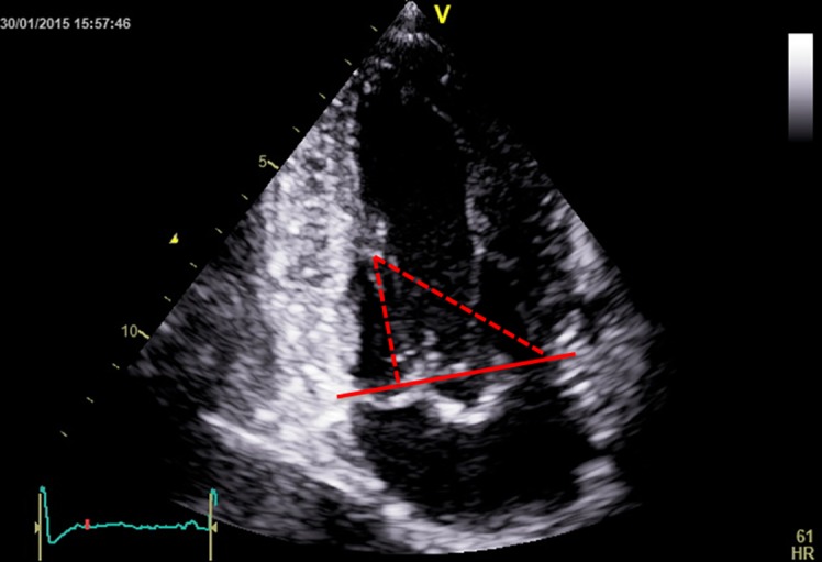
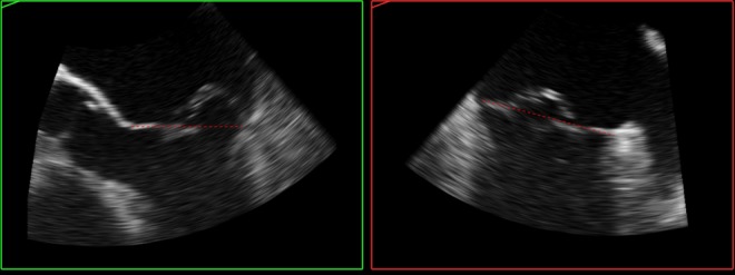
Papillary muscle to annulus plane (red continuous line) distances (red dotted lines).
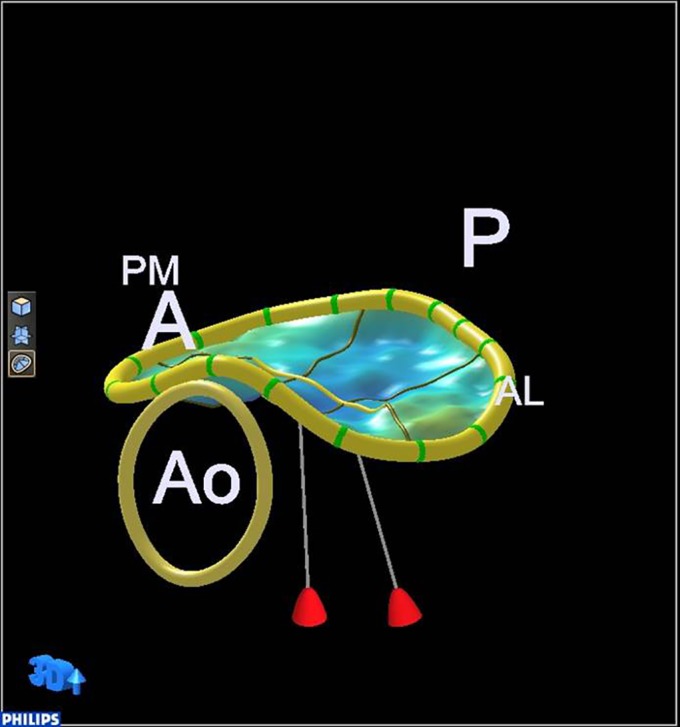
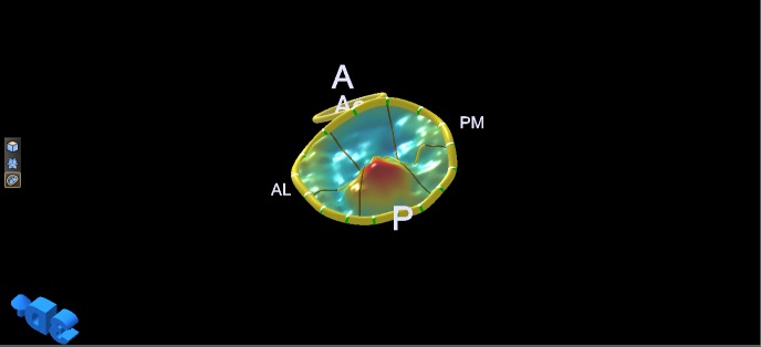
Normal annulus saddle shape. A, anterior; P, posterior; Ao, aortic valve; PM, postero-medial; AL, antero-lateral.
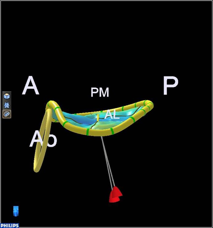
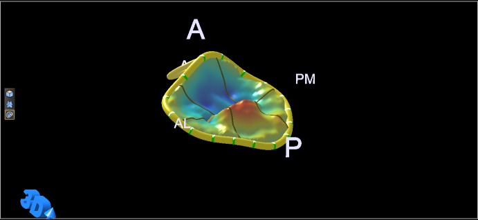
Normal annulus saddle and relative position of the papillary muscle tips. A, anterior; P, posterior; Ao, aortic valve; PM, postero-medial; AL, antero-lateral.
Predominant P2 prolapse.
Lone P2 prolapse.
3D model of the valve in Fig. 5 (lone P2 prolapse). A, anterior; P, posterior; Ao, aortic valve; PM, postero-medial; AL, antero-lateral.
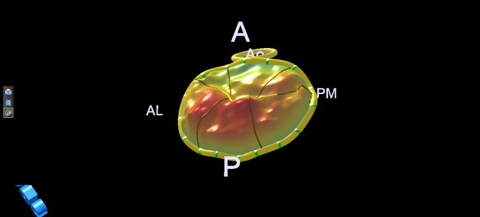
3D model of the valve in Fig. 4 (predominant P2 but also more extensive prolapse of the posterior leaflet and commissures). A, anterior; P, posterior; Ao, aortic valve; PM, postero-medial; AL, antero-lateral.
3D model of Barlow valve with extensive prolapse.

The 3D model from Fig. 8 displayed to reveal leaflet atrialisation.
TOE 2D images (MPR) derived from the 3D dataset at Fig. 5 demonstrating single scallop prolapse and otherwise thin, normal leaflets not billowing above the annulus plane (red dotted line).
TOE 2D images (MPR) derived from the 3D dataset of the valve from Fig. 8 for an alternative demonstration of leaflet atrialisation, with extensive billowing above the annulus plane (red dotted line).
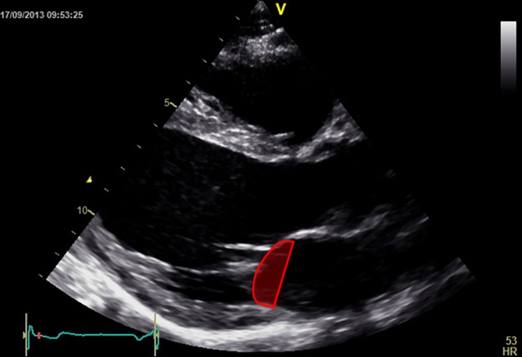
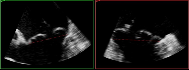
Tenting area.
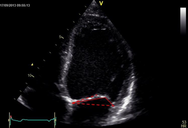
Tethering angles in valve with bent anterior leaflet. Red dotted lines define the annulus plane and the plane of the proximal anterior leaflet, and red continuous lines define the distal anterior leaflet and the posterior leaflet.
Similar articles
-
Automated quantification of mitral valve anatomy using anatomical intelligence in three-dimensional echocardiography.Int J Cardiol. 2015 Nov 15;199:232-8. doi: 10.1016/j.ijcard.2015.07.018. Epub 2015 Jul 11. Int J Cardiol. 2015. PMID: 26209825
-
Three-Dimensional Echocardiography: Advancements in Qualitative and Quantitative Analyses of Mitral Valve Morphology in Mitral Valve Prolapse.J Cardiovasc Echogr. 2014 Jan-Mar;24(1):1-9. doi: 10.4103/2211-4122.131985. J Cardiovasc Echogr. 2014. PMID: 28465897 Free PMC article. Review.
-
Contemporary imaging of normal mitral valve anatomy and function.Curr Opin Cardiol. 2012 Sep;27(5):455-64. doi: 10.1097/HCO.0b013e328354d7b5. Curr Opin Cardiol. 2012. PMID: 22820102 Review.
-
Quantification of mitral valve anatomy by three-dimensional transesophageal echocardiography in mitral valve prolapse predicts surgical anatomy and the complexity of mitral valve repair.J Am Soc Echocardiogr. 2012 Jul;25(7):758-65. doi: 10.1016/j.echo.2012.03.010. Epub 2012 Apr 24. J Am Soc Echocardiogr. 2012. PMID: 22537396
-
Relation of mitral valve morphology to surgical repair results in patients with mitral valve prolapse: A three-dimensional transesophageal echocardiography study.Echocardiography. 2018 Sep;35(9):1342-1350. doi: 10.1111/echo.14048. Epub 2018 Jun 19. Echocardiography. 2018. PMID: 29920772
Cited by
-
Papillary muscles of left ventricle-Morphological variations and it's clinical relevance.Indian Heart J. 2018 Nov-Dec;70(6):894-900. doi: 10.1016/j.ihj.2017.12.003. Epub 2017 Dec 11. Indian Heart J. 2018. PMID: 30580862 Free PMC article.
-
Arrhythmic Mitral Valve Prolapse: Introducing an Era of Multimodality Imaging-Based Diagnosis and Risk Stratification.Diagnostics (Basel). 2021 Mar 8;11(3):467. doi: 10.3390/diagnostics11030467. Diagnostics (Basel). 2021. PMID: 33800155 Free PMC article. Review.
-
Two and Three-Dimensional Echocardiography in Primary Mitral Regurgitation: Practical Hints to Optimize the Surgical Planning.Front Cardiovasc Med. 2021 Jul 8;8:706165. doi: 10.3389/fcvm.2021.706165. eCollection 2021. Front Cardiovasc Med. 2021. PMID: 34307510 Free PMC article. Review.
-
Imaging assessment of mitral and aortic regurgitation: current state of the art.Heart. 2020 Nov;106(22):1769-1776. doi: 10.1136/heartjnl-2019-316216. Epub 2020 Aug 17. Heart. 2020. PMID: 32817242 Free PMC article. Review. No abstract available.
-
The Role of 2D and 3D Echo in Mitral Stenosis.J Cardiovasc Dev Dis. 2021 Dec 3;8(12):171. doi: 10.3390/jcdd8120171. J Cardiovasc Dev Dis. 2021. PMID: 34940526 Free PMC article. Review.
References
-
- Lang RM Salgo IS Anyanwu AC Adams DH The road to mitral valve repair with live 3D transesophageal echocardiography Medicamundi 52 2008. 37–42.
-
- Shanks M Delgado V Ng ACT van der Kley F Schuijf JD Boersma E van de Veire NRL Nucifora G Bertini M de Roos A et al. Mitral valve morphology assessment: three-dimensional transesophageal echocardiography versus computed tomography Annals of Thoracic Surgery 90 2010. 1922–1929.10.1016/j.athoracsur.2010.06.116 - DOI - PubMed
Publication types
LinkOut - more resources
Full Text Sources
