

Neural correlates of abnormal sensory discrimination in laryngeal dystonia
- PMID: 26693398
- PMCID: PMC4660380
- DOI: 10.1016/j.nicl.2015.10.016
Neural correlates of abnormal sensory discrimination in laryngeal dystonia
Abstract
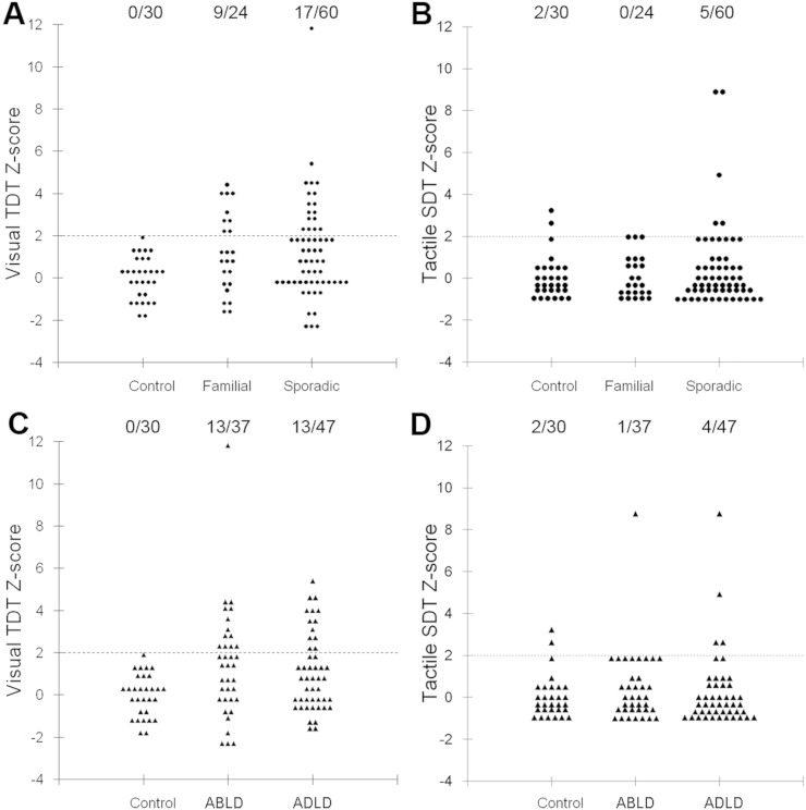
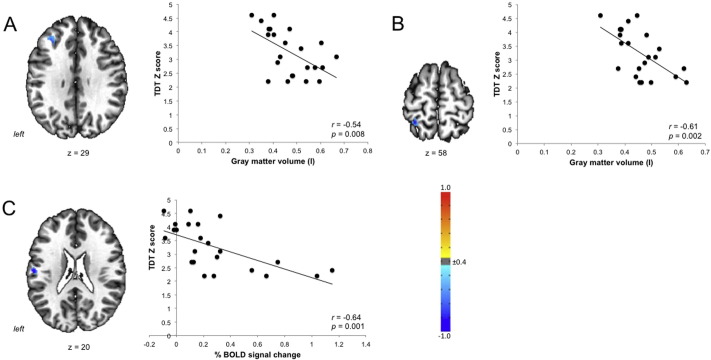
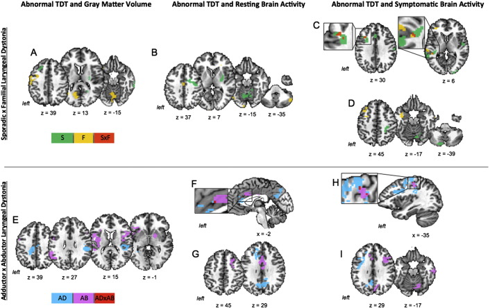
Aberrant sensory processing plays a fundamental role in the pathophysiology of dystonia; however, its underpinning neural mechanisms in relation to dystonia phenotype and genotype remain unclear. We examined temporal and spatial discrimination thresholds in patients with isolated laryngeal form of dystonia (LD), who exhibited different clinical phenotypes (adductor vs. abductor forms) and potentially different genotypes (sporadic vs. familial forms). We correlated our behavioral findings with the brain gray matter volume and functional activity during resting and symptomatic speech production. We found that temporal but not spatial discrimination was significantly altered across all forms of LD, with higher frequency of abnormalities seen in familial than sporadic patients. Common neural correlates of abnormal temporal discrimination across all forms were found with structural and functional changes in the middle frontal and primary somatosensory cortices. In addition, patients with familial LD had greater cerebellar involvement in processing of altered temporal discrimination, whereas sporadic LD patients had greater recruitment of the putamen and sensorimotor cortex. Based on the clinical phenotype, adductor form-specific correlations between abnormal discrimination and brain changes were found in the frontal cortex, whereas abductor form-specific correlations were observed in the cerebellum and putamen. Our behavioral and neuroimaging findings outline the relationship of abnormal sensory discrimination with the phenotype and genotype of isolated LD, suggesting the presence of potentially divergent pathophysiological pathways underlying different manifestations of this disorder.
Keywords: Brain imaging; Endophenotype; Sensory processing.
Figures
References
-
- Aglioti S.M., Fiorio M., Forster B., Tinazzi M. Temporal discrimination of cross-modal and unimodal stimuli in generalized dystonia. Neurology. 2003;60:782–785. - PubMed
-
- Ali S.O., Thomassen M., Schulz G.M., Hosey L.A., Varga M., Ludlow C.L., Braun A.R. Alterations in CNS activity induced by botulinum toxin treatment in spasmodic dysphonia: an H215O PET study. J. Speech Lang. Hear. Res. 2006;49:1127–1146. - PubMed
-
- Alvarez J.A., Emory E. Executive function and the frontal lobes: a meta-analytic review. Neuropsychol. Rev. 2006;16:17–42. - PubMed
-
- Ashburner J. A fast diffeomorphic image registration algorithm. Neuroimage. 2007;38:95–113. - PubMed
Publication types
MeSH terms
Grants and funding
LinkOut - more resources
Full Text Sources
Other Literature Sources
Medical
Research Materials
