

Relationship between the anterior forebrain mesocircuit and the default mode network in the structural bases of disorders of consciousness
- PMID: 26693399
- PMCID: PMC4660379
- DOI: 10.1016/j.nicl.2015.11.004
Relationship between the anterior forebrain mesocircuit and the default mode network in the structural bases of disorders of consciousness
Abstract
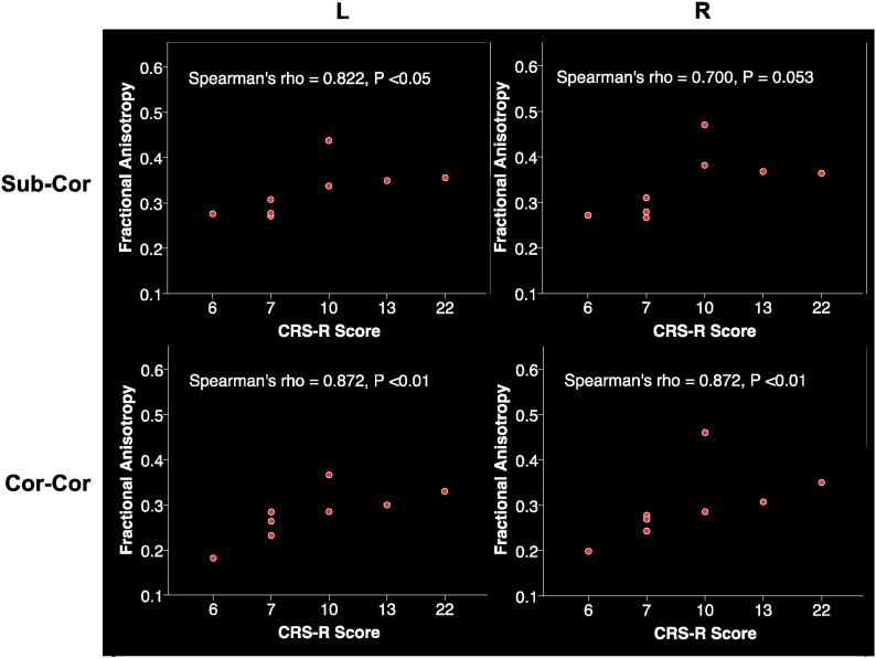
The specific neural bases of disorders of consciousness (DOC) are still not well understood. Some studies have suggested that functional and structural impairments in the default mode network may play a role in explaining these disorders. In contrast, others have proposed that dysfunctions in the anterior forebrain mesocircuit involving striatum, globus pallidus, and thalamus may be the main underlying mechanism. Here, we provide the first report of structural integrity of fiber tracts connecting the nodes of the mesocircuit and the default mode network in 8 patients with DOC. We found evidence of significant damage to subcortico-cortical and cortico-cortical fibers, which were more severe in vegetative state patients and correlated with clinical severity as determined by Coma Recovery Scale-Revised (CRS-R) scores. In contrast, fiber tracts interconnecting subcortical nodes were not significantly impaired. Lastly, we found significant damage in all fiber tracts connecting the precuneus with cortical and subcortical areas. Our results suggest a strong relationship between the default mode network - and most importantly the precuneus - and the anterior forebrain mesocircuit in the neural basis of the DOC.
Keywords: Anterior forebrain mesocircuit; Basal ganglia; DTI; Default mode network; Disorders of consciousness; Hypoxic–ischemic brain injury; Minimally conscious state; Precuneus; Thalamus; Tractography; Traumatic brain injury; Vegetative state; White matter.
Figures


References
-
- Adams J.H., Graham D.I., Murray L.S., Scott G. Diffuse axonal injury due to nonmissile head injury in humans: an analysis of 45 cases. Ann. Neurol. 1982;12:557–563. - PubMed
-
- Adams J.H., Graham D.I., Jennett B. The neuropathology of the vegetative state after an acute brain insult. Brain. 2000;123:1327–1338. - PubMed
-
- Alexander G.E., Crutcher M.D. Functional architecture of basal ganglia circuits: neural substrates of parallel processing. Trends Neurosci. 1990;13:266–271. - PubMed
-
- Alexander G.E., DeLong M.R., Strick P.L. Parallel organization of functionally segregated circuits linking basal ganglia and cortex. Annu. Rev. Neurosci. 1986;9:357–381. - PubMed
-
- Behrens T.E., Woolrich M.W., Jenkinson M., Johansen-Berg H., Nunes R.G., Clare S., Matthews P.M., Brady J.M., Smith S.M. Characterization and propagation of uncertainty in diffusion-weighted MR imaging. Magn. Reson. Med. 2003;50:1077–1088. - PubMed
Publication types
MeSH terms
Grants and funding
LinkOut - more resources
Full Text Sources
Other Literature Sources
