

Periventricular white matter abnormalities and restricted repetitive behavior in autism spectrum disorder
- PMID: 26693400
- PMCID: PMC4660377
- DOI: 10.1016/j.nicl.2015.10.017
Periventricular white matter abnormalities and restricted repetitive behavior in autism spectrum disorder
Abstract
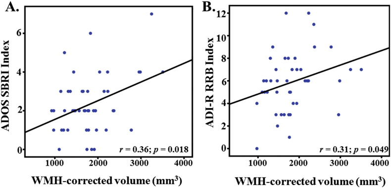
Malformations of cortical development are found at higher rates in autism spectrum disorder (ASD) than in healthy controls on postmortem neuropathological evaluation but are more variably observed on visual review of in-vivo MRI brain scans. This may be due to the visually elusive nature of many malformations on MRI. Here, we utilize a quantitative approach to determine whether a volumetric measure of heterotopic gray matter in the white matter is elevated in people with ASD, relative to typically developing controls (TDC). Data from a primary sample of 48 children/young adults with ASD and 48 age-, and gender-matched TDCs, selected from the Autism Brain Imaging Data Exchange (ABIDE) open-access database, were analyzed to compare groups on (1) blinded review of high-resolution T1-weighted research sequences; and (2) quantitative measurement of white matter hypointensity (WMH) volume calculated from the same T1-weighted scans. Groupwise WMH volume comparisons were repeated in an independent, multi-site sample (80 ASD/80 TDC), also selected from ABIDE. Visual review resulted in equivalent proportions of imaging abnormalities in the ASD and TDC group. However, quantitative analysis revealed elevated periventricular and deep subcortical WMH volumes in ASD. This finding was replicated in the independent, multi-site sample. Periventricular WMH volume was not associated with age but was associated with greater restricted repetitive behaviors on both parent-reported and clinician-rated assessment inventories. Thus, findings demonstrate that periventricular WMH volume is elevated in ASD and associated with a higher degree of repetitive behaviors and restricted interests. Although the etiology of focal WMH clusters is unknown, the absence of age effects suggests that they may reflect a static anomaly.
Keywords: Autism spectrum disorder; Magnetic resonance imaging; Malformations of cortical development; Periventricular heterotopia; Restricted repetitive behaviors; White matter hypointensities.
Figures

Similar articles
-
Longitudinal volumetric brain changes in autism spectrum disorder ages 6-35 years.Autism Res. 2015 Feb;8(1):82-93. doi: 10.1002/aur.1427. Epub 2014 Nov 7. Autism Res. 2015. PMID: 25381736 Free PMC article.
-
Anatomical likelihood estimation meta-analysis of grey and white matter anomalies in autism spectrum disorders.Neuroimage Clin. 2014 Nov 18;7:525-36. doi: 10.1016/j.nicl.2014.11.004. eCollection 2015. Neuroimage Clin. 2014. PMID: 25844306 Free PMC article.
-
The effect of gender on the neuroanatomy of children with autism spectrum disorders: a support vector machine case-control study.Mol Autism. 2016 Jan 19;7:5. doi: 10.1186/s13229-015-0067-3. eCollection 2016. Mol Autism. 2016. PMID: 26788282 Free PMC article.
-
Structural MRI biomarkers of shared pathogenesis in autism spectrum disorder and epilepsy.Epilepsy Behav. 2015 Jun;47:172-82. doi: 10.1016/j.yebeh.2015.02.017. Epub 2015 Mar 24. Epilepsy Behav. 2015. PMID: 25812936 Review.
-
Neurodevelopmental sequelae associated with gray and white matter changes and their cellular basis: A comparison between Autism Spectrum Disorder, ADHD and dyslexia.Int J Dev Neurosci. 2015 Nov;46:132-43. doi: 10.1016/j.ijdevneu.2015.02.007. Epub 2015 Aug 11. Int J Dev Neurosci. 2015. PMID: 26456538 Review.
Cited by
-
Behavioral Phenotyping for Autism Spectrum Disorders in Mice.Curr Protoc Toxicol. 2017 May 2;72:11.22.1-11.22.21. doi: 10.1002/cptx.19. Curr Protoc Toxicol. 2017. PMID: 28463420 Free PMC article.
-
Description of Motor Stereotypies in Adolescents and Adults With Autistic Spectrum Disorder.Actas Esp Psiquiatr. 2025 Aug;53(4):715-729. doi: 10.62641/aep.v53i4.1917. Actas Esp Psiquiatr. 2025. PMID: 40791045 Free PMC article.
-
Autism spectrum disorder: neuropathology and animal models.Acta Neuropathol. 2017 Oct;134(4):537-566. doi: 10.1007/s00401-017-1736-4. Epub 2017 Jun 5. Acta Neuropathol. 2017. PMID: 28584888 Free PMC article. Review.
-
Dysfunction of the Auditory Brainstem as a Neurophysiology Subtype of Autism Spectrum Disorder.Front Neurosci. 2021 Mar 17;15:637079. doi: 10.3389/fnins.2021.637079. eCollection 2021. Front Neurosci. 2021. PMID: 33815042 Free PMC article.
-
The neural circuitry of restricted repetitive behavior: Magnetic resonance imaging in neurodevelopmental disorders and animal models.Neurosci Biobehav Rev. 2018 Sep;92:152-171. doi: 10.1016/j.neubiorev.2018.05.022. Epub 2018 May 23. Neurosci Biobehav Rev. 2018. PMID: 29802854 Free PMC article. Review.
References
-
- American Psychiatric Association . 5th ed. American Psychiatric Publishing; Arlington, VA: 2013. Diagnostic and Statistical Manual of Mental Disorders.
-
- Bergeron J.D., Deslauriers J., Grignon S., Fortier L.C., Lepage M., Stroh T. White matter injury and autistic-like behavior predominantly affecting male rat offspring exposed to group B streptococcal maternal inflammation. Dev. Neurosci. 2013;35:504–515. - PubMed
-
- Bernasconi A., Bernasconi N., Bernhardt B.C., Schrader D. Advances in MRI for ‘cryptogenic’ epilepsies. Nat. Rev. Neurol. 2011;7:99–108. - PubMed
Publication types
MeSH terms
Grants and funding
LinkOut - more resources
Full Text Sources
Other Literature Sources
Medical
