

Basal Cell Adenoma of Parotid Gland: Case Report and Review of Literature
- PMID: 26693465
- PMCID: PMC4678265
- DOI: 10.1007/s12070-015-0865-0
Basal Cell Adenoma of Parotid Gland: Case Report and Review of Literature
Abstract
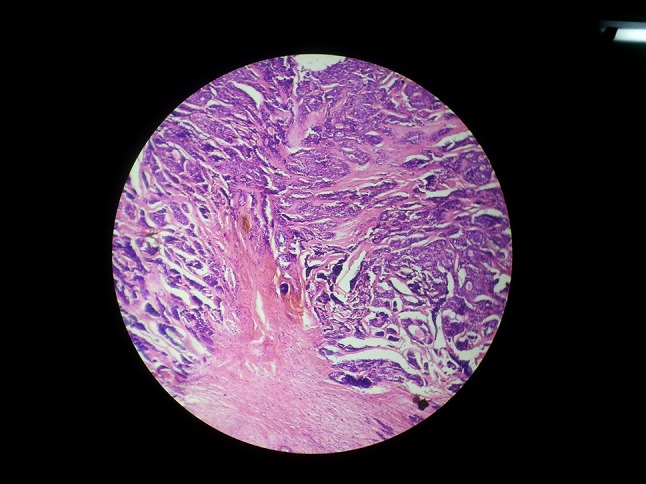
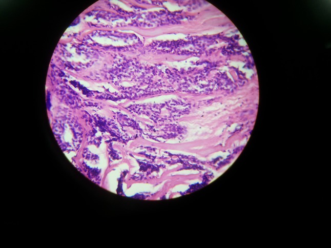
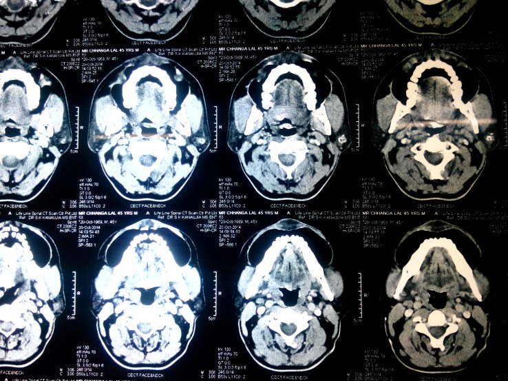
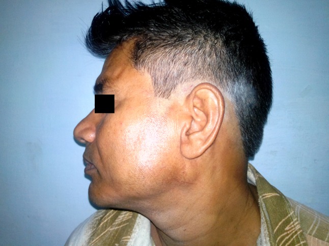
Basal cell adenoma (BCA) of the salivary gland is a rare neoplasm consists of a monomorphic population of basaloid epithelial cells, and it accounts for approximately 1-2 % of all salivary gland tumors. Its most frequent location is the parotid gland. It usually appears as a firm and mobile slow-growing mass. Histologically, isomorphic cells in nests and interlaced trabecules with a prominent basal membrane are observed. In contrast to pleomorphic adenoma, it tends to be multiple and its recurrence rate after surgical excision is high. Due to prognostic implications, differential diagnosis with basal cell adenocarcinoma, adenoid cystic carcinoma and basaloid squamous cell carcinoma is mandatory. We report a case of BCA of the parotid gland. We also review the literature and discuss the diagnosis and management of this rare entity.
Keywords: Basal cell adenoma; Parotid gland; Salivary gland tumors.
Figures

Similar articles
-
Basal cell adenoma of parotid gland: two case reports and literature review.Arch Craniofac Surg. 2023 Aug;24(4):179-184. doi: 10.7181/acfs.2023.00227. Epub 2023 Aug 20. Arch Craniofac Surg. 2023. PMID: 37654238 Free PMC article.
-
Basal cell adenoma of the parotid gland. Case report and review of the literature.Med Oral Patol Oral Cir Bucal. 2006 Mar 1;11(2):E206-9. Med Oral Patol Oral Cir Bucal. 2006. PMID: 16505803 Review. English, Spanish.
-
Basal Cell Adenoma of the Parotid Gland.Kathmandu Univ Med J (KUMJ). 2020 Oct.-Dec.;18(72):434-436. Kathmandu Univ Med J (KUMJ). 2020. PMID: 34165108 Review.
-
Basal cell adenoma in a relatively rare site.J Oral Maxillofac Pathol. 2009 Jul;13(2):101-4. doi: 10.4103/0973-029X.57680. J Oral Maxillofac Pathol. 2009. PMID: 21887012 Free PMC article.
-
Basal cell adenoma with extensive squamous metaplasia and cellular atypia: a case report with cytohistopathological correlation and review of the literature.Diagn Cytopathol. 2012 Jan;40(1):48-55. doi: 10.1002/dc.21584. Epub 2010 Dec 3. Diagn Cytopathol. 2012. PMID: 22180238 Review.
Cited by
-
Solitary juvenile xanthogranuloma in the parotid gland.Radiol Case Rep. 2023 Mar 3;18(5):1784-1787. doi: 10.1016/j.radcr.2023.01.103. eCollection 2023 May. Radiol Case Rep. 2023. PMID: 36923389 Free PMC article.
-
Basal cell adenoma of parotid gland: two case reports and literature review.Arch Craniofac Surg. 2023 Aug;24(4):179-184. doi: 10.7181/acfs.2023.00227. Epub 2023 Aug 20. Arch Craniofac Surg. 2023. PMID: 37654238 Free PMC article.
-
Basal Cell Adenoma of the Parotid Gland: A Rare Disease Entity.Cureus. 2023 Mar 29;15(3):e36841. doi: 10.7759/cureus.36841. eCollection 2023 Mar. Cureus. 2023. PMID: 37123726 Free PMC article.
-
Basal cell adenoma and basal cell adenocarcinoma of the parotid gland: clinical findings and surgical outcomes in a single-institution study.World J Surg Oncol. 2024 Apr 18;22(1):102. doi: 10.1186/s12957-024-03378-6. World J Surg Oncol. 2024. PMID: 38637826 Free PMC article.
-
Cytodiagnosis of Basal Cell Adenoma of Parotid Gland: A Rare Case Report.Cureus. 2023 Mar 27;15(3):e36767. doi: 10.7759/cureus.36767. eCollection 2023 Mar. Cureus. 2023. PMID: 37123745 Free PMC article.
References
-
- Gök Ü, Sakallıoğlu Ö, Dağlı AF. Basal cell adenoma of the parotide gland: case report. Fırat Tıp Dergisi. 2006;11:150–152.
-
- Siefert G, Sobin LH (1991) Histological classification of salivary gland tumours. In: World Health Organization, International histological classification of tumours. Springer, Berlin
Publication types
LinkOut - more resources
Full Text Sources
Other Literature Sources